Comparative Study
. 2011 Jul;1(3):280-94.
doi: 10.1016/j.dcn.2011.02.002.

Enhanced neural responses to rule violation in children with autism: a comparison to social exclusion

Affiliations
Comparative Study

Enhanced neural responses to rule violation in children with autism: a comparison to social exclusion

Danielle Z Bolling et al. Dev Cogn Neurosci. 2011 Jul.

Abstract

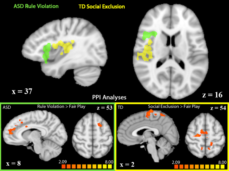
The present study aimed to explore the neural correlates of two characteristic deficits in autism spectrum disorders (ASD); social impairment and restricted, repetitive behavior patterns. To this end, we used comparable experiences of social exclusion and rule violation to probe potentially atypical neural networks in ASD. In children and adolescents with and without ASD, we used the interactive ball-toss game (Cyberball) to elicit social exclusion and a comparable game (Cybershape) to elicit a non-exclusive rule violation. Using functional magnetic resonance imaging (fMRI), we identified group differences in brain responses to social exclusion and rule violation. Though both groups reported equal distress following exclusion, the right insula and ventral anterior cingulate cortex were hypoactive during exclusion in children with ASD. In rule violation, right insula and dorsal prefrontal cortex were hyperactive in ASD. Right insula showed a dissociation in activation; it was hypoactive to social exclusion and hyperactive to rule violation in the ASD group. Further probed, different regions of right insula were modulated in each game, highlighting differences in regional specificity for which subsequent analyses revealed differences in patterns of functional connectivity. These results demonstrate neurobiological differences in processing social exclusion and rule violation in children with ASD.

Keywords: Autism Spectrum Disorder; Functional Magnetic Resonance Imaging; Right Insula; Rule Violation; Social Exclusion.


Figures

Fig. 1
Fig. 1
Top: whole-brain comparison of social exclusion and fair play in TD (n = 21; left) and ASD participants (n = 16; right). Regions in orange showed greater activation in social exclusion compared to fair play. Regions in blue showed greater activation in fair play compared to social exclusion (p < 0.05, k = 34). Bottom: whole-brain comparison of rule violation and fair play in TD (n = 19; left) and ASD participants (n = 21; right). Regions in orange showed greater activation in rule violation compared to fair play. Regions in blue showed greater activation in fair play compared to rule violation (p < 0.05, k = 34). Activations are displayed on a Talairach-transformed template brain in radiological orientation. (For interpretation of the references to color in this figure legend, the reader is referred to the web version of the article.)
Fig. 2
Fig. 2
Regions showing significant Group × Condition interactions in Cyberball (top) and Cybershape (bottom; p < 0.05, k = 20). Bar graphs depict beta value differences (social exclusion–fair play or rule violation–fair play) in regions of interaction. Regions described are not inclusive of all regions showing a significant interaction. * Denotes similar patterns of interaction bilaterally. Activations are displayed on a Talairach-transformed template brain in radiological orientation.
Fig. 3
Fig. 3
Group × Game interaction in a structurally defined region of right insula (left panel). Beta value differences in each group were graphed for Cyberball (social exclusion–fair play; middle panel) and Cybershape (rule violation–fair play; right panel). This Group × Game interaction was significant at a p = 0.007.
Fig. 4
Fig. 4
PPI analyses in regions showing significant activation in TD children during social exclusion (shown in yellow) or in ASD children during rule violation (green). Seed regions for the PPI analyses are depicted in the top panel. Regions showing increased functional connectivity to seed regions during social exclusion (right) or rule violation (left) are depicted in the bottom panel (p < 0.05, k = 34). Activations are displayed on a Talairach-transformed template brain in radiological orientation. (For interpretation of the references to color in this figure legend, the reader is referred to the web version of the article.)
Fig. 5
Fig. 5
Whole brain correlation analysis in Cyberball with all participants. Regions in blue showed a negative correlation with Social Responsiveness Scale (SRS) scores in the contrast social exclusion > fair play. These regions were more active, the more socially responsive the participant (p < 0.01, k = 4). Activations are displayed on a Talairach-transformed template brain in radiological orientation. (For interpretation of the references to color in this figure legend, the reader is referred to the web version of the article.)

Comment in

References

    1. American Psychiatric Association . Diagnostic and Statistical Manual of Mental Disorders, 4th ed. (DSM-IV-TR) American Psychiatric Association; Washington, DC: 2000. Pervasive developmental disorders.
    1. Andari E., Duhamel J.-R., Zalla T., Herbrecht E., Leboyer M., Sirigu A. Promoting social behavior with oxytocin in high-functioning autism spectrum disorders. Proc. Natl. Acad. Sci. U.S.A. 2010;107:4389–4394. - PMC - PubMed
    1. Bolling D.Z., Pitskel N.B., Deen B., Crowley M.J., McPartland J.C., Mayes L.C., Pelphrey K.A. Dissociable brain mechanisms for processing social exclusion and rule violation. NeuroImage. 2011;54:2426–2471. - PMC - PubMed
    1. Brooks J.C.W., Nurmikko T.J., Bimson W.E., Singh K.D., Roberts N. fMRI of thermal pain: effects of stimulus laterality and attention. NeuroImage. 2002;15:293–301. - PubMed
    1. Buckner R.L., Andrews-Hanna J.R., Schachter D.L. The brain's default network: anatomy, function, and relevance to disease. Ann. N.Y. Acad. Sci. 2008;1124:1–38. - PubMed

Publication types

LinkOut - more resources