. 2012 Feb;69(4):611-27.
doi: 10.1007/s00018-011-0765-8. Epub 2011 Jul 9.

Hes-1 regulates the excitatory fate of neural progenitors through modulation of Tlx3 (HOX11L2) expression

Affiliations

Hes-1 regulates the excitatory fate of neural progenitors through modulation of Tlx3 (HOX11L2) expression

Chandrasekharan Lalitha Indulekha et al. Cell Mol Life Sci. 2012 Feb.

Abstract

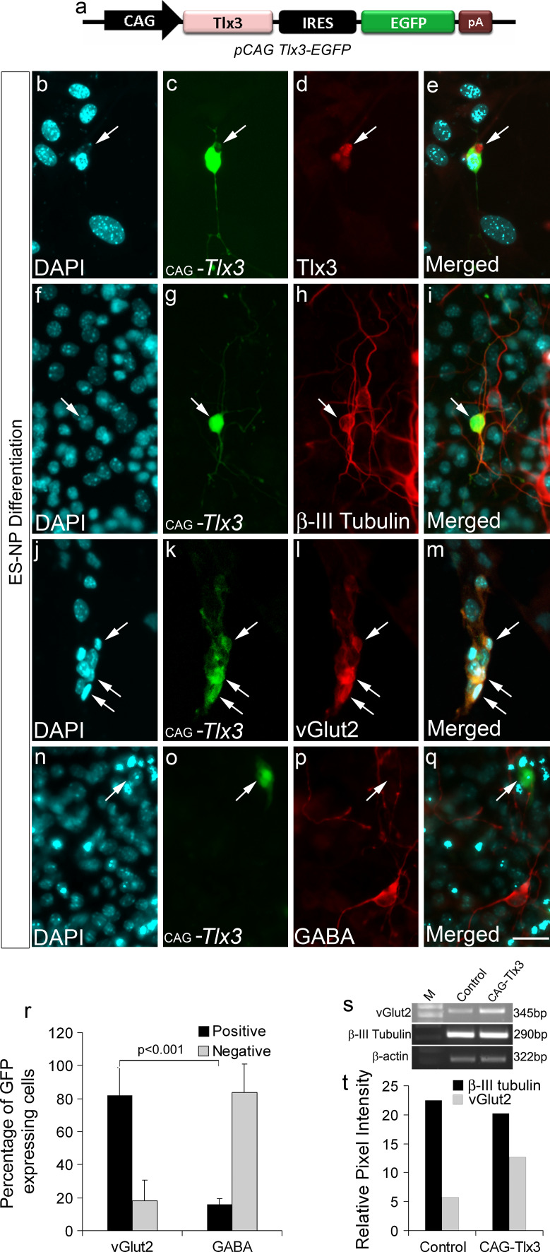
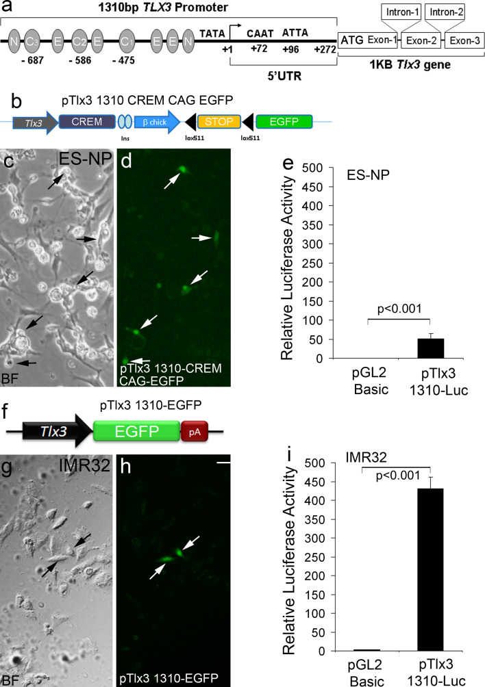
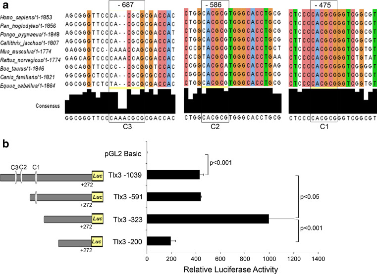
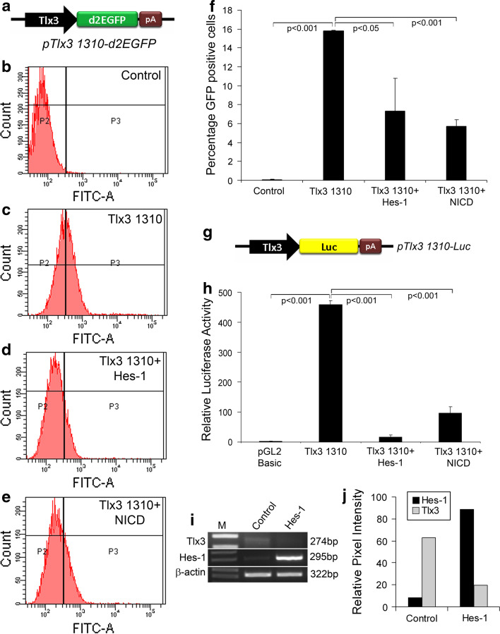
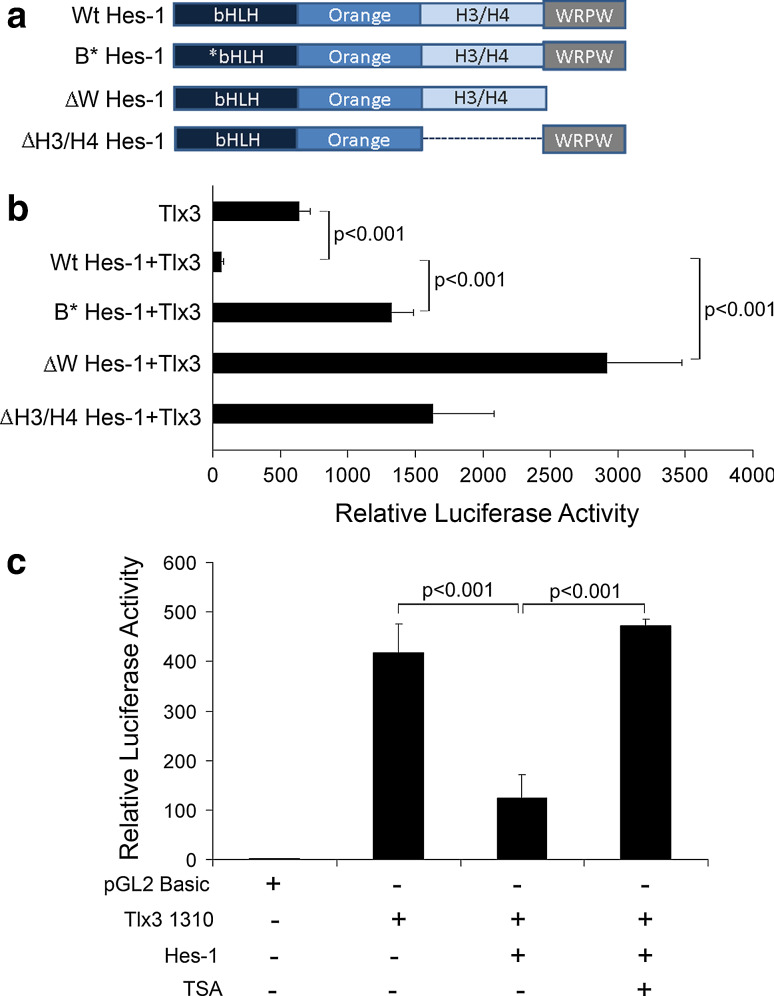
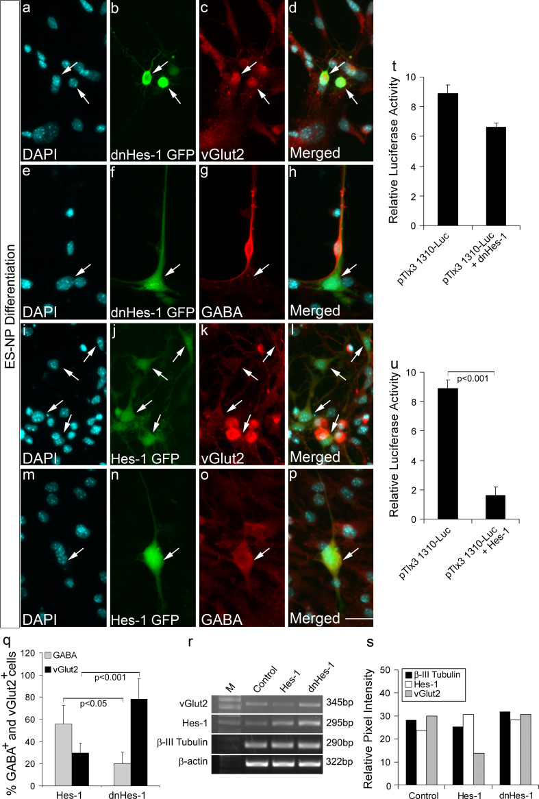
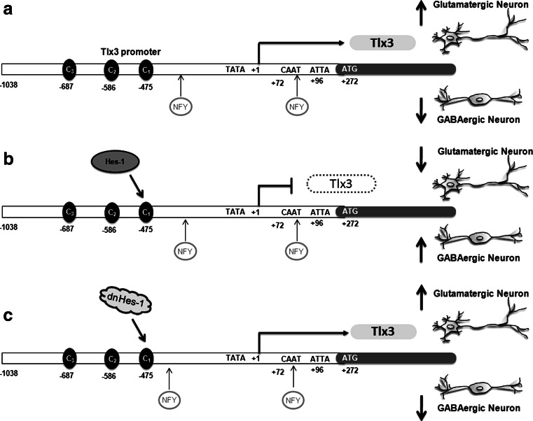
Tlx3 (HOX11L2) is regarded as one of the selector genes in excitatory versus inhibitory fate specification of neurons in distinct regions of the nervous system. Expression of Tlx3 in a post-mitotic immature neuron favors a glutamatergic over GABAergic fate. The factors that regulate Tlx3 have immense importance in the fate specification of glutamatergic neurons. Here, we have shown that Notch target gene, Hes-1, negatively regulates Tlx3 expression, resulting in decreased generation of glutamatergic neurons. Down-regulation of Hes-1 removed the inhibition on Tlx3 promoter, thus promoting glutamatergic differentiation. Promoter-protein interaction studies with truncated/mutated Hes-1 protein suggested that the co-repressor recruitment mediated through WRPW domain of Hes-1 has contributed to the repressive effect. Our results clearly demonstrate a new and unique role for canonical Notch signaling through Hes-1, in neurotransmitter/subtype fate specification of neurons in addition to its known functional role in proliferation/maintenance of neural progenitors.


Figures

Fig. 1
Fig. 1
Up-regulation of Tlx3 in ES-NPs promotes a glutamatergic fate: a Schematic of Tlx3 expression construct, where Tlx3 is expressed under the control of CAG promoter with IRES-EGFP so that the transfected cells will be expressing GFP. be Authenticity of Tlx3 expression construct was validated by anti-Tlx3 immunostaining which showed Tlx3 expression in transfected cells. fi Tlx3-expressing cells were positive for neuronal marker β-III tubulin, thus confirming their neuronal nature. jq Tlx3-expressing cells were vGlut2-positive and were negative for GABA. r Quantitative immunocytochemical analysis of vGlut2 and GABA in Tlx3 over-expressed cells showed a significantly high percentage (p < 0.001) of cells undergoing glutamatergic differentiation compared to GABAergic differentiation. st RT-PCR analysis showed increased vGlut2 expression in Tlx3 over-expressed ES-NPs. Data are expressed as mean ± SD of triplicates (n = 3) from three different experiments. Scale bar = 50 µM
Fig. 2
Fig. 2
Functional analysis of the Tlx3 promoter in ES-NPs and IMR32 cell lines: a Schematic of the Tlx3 promoter with different transcription factor-binding sites. b Tlx3-driven GFP expression cassette used to transfect ES-NPs. Since the level of expression of Tlx3 is low, we enhanced the visualization of Tlx3 expression using a Cre-lox construct, pTlx3 1310-CREM-CAG-EGFP. cd ES-NPs upon differentiation showed GFP expression indicating functional activity of the cloned Tlx3 promoter. e Luciferase activity in ES-NPs upon transient transfection of pTlx3-1310-Luc in differentiated ES-NPs showed a significant increase (p < 0.001) compared to control. f Schematic of Tlx3 promoter-GFP reporter construct used to study Tlx3 expression in the IMR32 cell line. gh Transient transfections with pTlx3-1310-EGFP construct showed GFP-positive cells, indicating Tlx3 expression in IMR32 cell line. i This was again confirmed by assaying Tlx3 promoter activity, which showed a significant increase (p < 0.001) in luciferase expression compared to the control. Significantly high promoter activation indicates a higher level of expression of Tlx3 in IMR32 cell line compared to ES-NPs. Data are expressed as mean ± SD of triplicates (n = 3) from three different experiments. Scale bar = 20 µM
Fig. 3
Fig. 3
Hes-1-binding C sites are involved in regulation of Tlx3 promoter: a C1, C2 and C3 sites showed 100, 66.6, and 55.5% consensus among different mammalian species. Consensus was analyzed using the software "Jalview version 2". b Tlx3 1310 promoter with all the three C sites showed a significant increase (p < 0.001) in luciferase activity compared to the control. Deletion of C2 and C3 sites did not show any significant reduction in luciferase activity, but deletion of the proximal C1 site (Del-323) significantly increased (p < 0.05) the luciferase activity indicating that this C-site is critical for the repression of the Tlx3 promoter by Hes-1. Further deletion of 123-bp (Del-200) resulted in a significant reduction (p < 0.001) in activity compared to Del-323, indicating that this 123-bp region might be involved in the possible activation of Tlx3. Data are expressed as mean ± SD of triplicates (n = 3) from three different experiments
Fig. 4
Fig. 4
Hes-1 acts as a repressor of Tlx3 promoter: a Schematic of Tlx3 promoter-driven d2EGFP having a half-life of 2 h so that the change in Tlx3 expression is accurately reflected by the GFP expression. be FACS analysis of IMR32 cells transfected with pTlx3 1310-d2GFP in the presence or absence of Hes-1 or NICD. f Graph depicting the percentage of GFP-positive cells obtained with FACS analysis. Transfection with pTlx3-1310-d2EGFP alone significantly increased (p < 0.001) the percentage of GFP-expressing cells, whereas co-transfection with Hes-1 and NICD significantly reduced (p < 0.05 and p < 0.001, respectively) the percentage of GFP-expressing cells. g Schematic of Tlx3 promoter-driven luciferase reporter system. h Luciferase assay of Tlx3 promoter showed a significant increase (p < 0.001) in activity compared to control. Expression of Hes-1 alone with Tlx3 promoter showed a significant decrease (p < 0.001) in Tlx3 promoter activity. ij RT-PCR analysis of Tlx3 in IMR32 cells showed a reduction in Tlx3 expression upon Hes-1 transfection. Data are expressed as mean ± SD of triplicates (n = 3) from three different experiments
Fig. 5
Fig. 5
WRPW domain mediated-protein interaction along with DNA- binding and histone deacetylase activity is involved in regulation of Tlx3 promoter: a Schematic representation of different functional domains of Wt type Hes-1 and different truncated/mutated Hes-1 proteins used in this study. B*Hes-1 represents basic domain-mutated Hes-1 and thus is unable to bind target DNA. However, all of the other domains are intact so that it can interact with other proteins and is able to recruit co-repressor proteins and can dimerize with other bHLH factors. ∆W Hes-1 represents WRPW domain truncation with deletion of extreme C-terminal four amino acids. ∆W Hes-1 is able to carry out all functions of Hes-1 except co-repressor recruitment. ∆H3/H4 Hes-1 indicates H3/H4 domain truncated Hes-1, where protein–protein interactions may be affected. b Luciferase activity was measured with co-transfection of the above-mentioned Hes-1 constructs along with pTlx3 1310-Luc to study the contribution of different functional domains of Hes-1 in Tlx3 promoter regulation. The Wt type Hes-1 significantly repressed (p < 0.001) Tlx3 promoter activity, whereas B*Hes-1 significantly reduced (p < 0.001) the repression compared to Wt type, indicating a function role of DNA binding in Tlx3 promoter regulation. Again, ∆W Hes-1 with WRPW domain truncation showed a significant activation/de-repression (p < 0.001) of the promoter, indicating an active co-repressor recruitment role in Tlx3 promoter regulation. ∆H3/H4 domain truncated Hes-1 also did not show any repression indicating functional protein interactions even with co-repressors in Hes-1-mediated repression. c Treatment with deacetylase inhibitor, Trichostatin A (TSA) resulted in the abolishment of inhibition caused by Hes-1. These results indicated that histone deacetylase activity is one of the mechanisms through which Hes-1 represses the Tlx3 promoter
Fig. 6
Fig. 6
Functional role of Hes-1 in glutamatergic versus GABAergic fate specification of neural progenitors: dnHes-1 GFP and Hes-1 GFP constructs were transfected in ES-NPs and were allowed to differentiate further for 6 days. ah dnHes-1 GFP-transfected cells showed robust glutamatergic differentiation as evidenced by expression of vGlut2 and were negative for GABA. ip The majority of Hes-1 GFP-transfected cells were negative for vGlut2 and were positive for GABA. These results show that down-regulation of Hes-1 removes the repression on Tlx3 promoter leading to robust glutamatergic differentiation. q Graph represents quantitative analysis of percentage of glutamatergic and GABAergic neurons differentiated in the presence of Hes-1 and dnHes-1. ES-NPs transfected with dnHes-1 showed a significant increase (p < 0.001) in vGlut2-positive cells compared to those transfected with Hes-1, whereas ES-NPs transfected with Hes-1 showed a significant increase (p < 0.05) in GABA-positive cells compared to those transfected with dnHes-1. rs RT-PCR analysis showed decreased vGlut2 expression in the presence of Hes-1 compared to the control and enhanced Hes-1 expression in the presence of dnHes-1 in differentiated ES-NPs. The expression of Hes-1 was increased by transfection of Hes-1 and dnHes-1 since our Hes-1 primer was designed to amplify both Hes-1 and dnHes-1, there by confirming its up-regulation in the transfected cells. Expression of β-III tubulin in all the conditions indicates robust neuronal differentiation. t Luciferase activity in ES-NPs transfected with dnHes-1 does not show a significant decrease in Tlx3 expression compared to the control. The ES-NPs used for this experiment had stable integration of pTlx3 1310-Luc. u Luciferase activity in ES-NPs transfected with Hes-1 showed a significant decrease (p < 0.001) in Tlx3 expression compared to control. In panel ar the progenitors were transfected with Hes-1 and dnHes-1 alone, whereas in panels tu the progenitors having stable integration of Tlx3 promoter-Luciferase construct were transfected with Hes-1 and dnHes-1 vectors. Data are expressed as mean ± SD of triplicates (n = 3) from three different experiments. Scale bar = 50 µM
Fig. 7
Fig. 7
Schematic showing the regulation of Tlx3 by Hes-1 during neuronal fate specification: a Expression of Tlx3 through NFY promotes glutamatergic fate over its complementary GABAergic fate. b Expression of Hes-1 represses Tlx3 by binding to the C1 site, thereby suppressing glutamatergic fate and increasing the GABAergic neurons. c Down-regulation of Hes-1 expression by dnHes-1 removes the repression on the Tlx3 promoter, thus enhancing the differentiation of glutamatergic neurons

Similar articles

Cited by

References

    1. Jan YN, Jan LY. HLH proteins, fly neurogenesis, and vertebrate myogenesis. Cell. 1993;75:827–830. doi: 10.1016/0092-8674(93)90525-U. - DOI - PubMed
    1. Jessell TM. Neuronal specification in the spinal cord: inductive signals and transcriptional codes. Nat Rev Genet. 2000;1:20–29. doi: 10.1038/35049541. - DOI - PubMed
    1. Kintner C. Neurogenesis in embryos and in adult neural stem cells. J Neurosci. 2002;22:639–643. - PMC - PubMed
    1. Ma Q. Transcriptional regulation of neuronal phenotype in mammals. J Physiol. 2006;575:379–387. doi: 10.1113/jphysiol.2006.113449. - DOI - PMC - PubMed
    1. Guillemot F. Spatial and temporal specification of neural fates by transcription factor codes. Development. 2007;134:3771–3780. doi: 10.1242/dev.006379. - DOI - PubMed

Publication types

MeSH terms