Review
. 2011 Dec;34(6):1183-97.
doi: 10.1007/s10545-011-9359-8. Epub 2011 Jul 9.

Cardiac disease in patients with mucopolysaccharidosis: presentation, diagnosis and management

Affiliations
Review

Cardiac disease in patients with mucopolysaccharidosis: presentation, diagnosis and management

Elizabeth A Braunlin et al. J Inherit Metab Dis. 2011 Dec.

Abstract

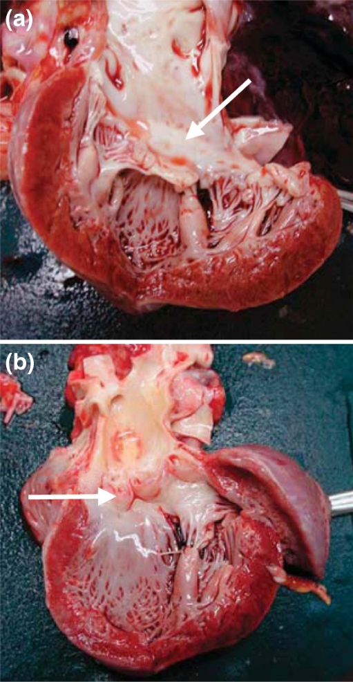
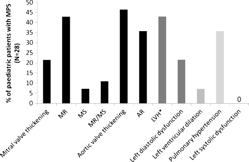
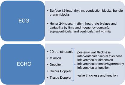
The mucopolysaccharidoses (MPSs) are inherited lysosomal storage disorders caused by the absence of functional enzymes that contribute to the degradation of glycosaminoglycans (GAGs). The progressive systemic deposition of GAGs results in multi-organ system dysfunction that varies with the particular GAG deposited and the specific enzyme mutation(s) present. Cardiac involvement has been reported in all MPS syndromes and is a common and early feature, particularly for those with MPS I, II, and VI. Cardiac valve thickening, dysfunction (more severe for left-sided than for right-sided valves), and hypertrophy are commonly present; conduction abnormalities, coronary artery and other vascular involvement may also occur. Cardiac disease emerges silently and contributes significantly to early mortality.The clinical examination of individuals with MPS is often difficult due to physical and, sometimes, intellectual patient limitations. The absence of precordial murmurs does not exclude the presence of cardiac disease. Echocardiography and electrocardiography are key diagnostic techniques for evaluation of valves, ventricular dimensions and function, which are recommended on a regular basis. The optimal technique for evaluation of coronary artery involvement remains unsettled.Standard medical and surgical techniques can be modified for MPS patients, and systemic therapies such as hematopoietic stem cell transplantation and enzyme replacement therapy (ERT) may alter overall disease progression with regression of ventricular hypertrophy and maintenance of ventricular function. Cardiac valve disease is usually unresponsive or, at best, stabilized, although ERT within the first few months of life may prevent valve involvement, a fact that emphasizes the importance of early diagnosis and treatment in MPS.


Figures

Fig. 1
Fig. 1
(a) Autopsy specimen from a female patient with Hurler syndrome at the age of 2.8 years showing (a) a dysplastic and thickened mitral valve (arrow) with a thickened subvalvular apparatus and parietal left ventricular hypertrophy and (b) a dysplastic aortic valve (arrow) (Fesslová et al. 2009) (copyright Fesslová et al. Cardiol Young 2009)
Fig. 2
Fig. 2
Echocardiography and Doppler interrogation of mitral (a) and aortic (b) valve in a 50-year-old male with MPS VI (a) Short-axis view of thickened mitral valve (arrow) in diastole (left). Colour Doppler demonstration of mitral regurgitation (right). (b) Short-axis view demonstrating thickened trileaflet aortic valve (arrow) in systole (left). Colour Doppler demonstrating turbulent systolic flow of aortic valve stenosis (right)
Fig. 3
Fig. 3
Echocardiographic evaluations in patients with MPS (2–14 years old, mean age 9 years; MPS I-II-III-IV-VI-VII) indicate that left valve lesions, left ventricular hypertrophy (LVH) and pulmonary hypertension are the most common cardiac findings (Leal et al. 2010). AR: aortic regurgitation; MR: mitral regurgitation; MS: mitral stenosis; MR/MS: both lesions; *LVH: septal and posterior wall hypertrophy of left ventricle
Fig. 4
Fig. 4
Epicardial right coronary artery from untreated 1-year-old male with rapidly progressing MPS I. Note diffuse myointimal proliferation (arrow). (Alcian blue stain)
Fig. 5
Fig. 5
Light microscopy of the mitral valve of a girl with MPS VI at 14 years of age, demonstrating enlarged and foamy appearing fibroblasts (arrows) (hematoxylin and eosin staining, magnification x 20)
Fig. 6
Fig. 6
Recommended diagnostic techniques and assessments to evaluate cardiac anatomy and function in patients with MPS at initial diagnosis and at regular intervals thereafter (every 1 to 2 years for MPS I and VI, or 1 to 3 years for MPS II) 2D: 2-dimensional; ECG: electrocardiography; ECHO: echocardiography

References

    1. Aguilar F, Nesser HJ, Faletra F, et al. Imaging modalities in valvular heart disease. Curr Cardiol Rep. 2008;10:98–103. doi: 10.1007/s11886-008-0018-0. - DOI - PubMed
    1. Antoniou T, Kirvassilis G, Tsourelis L, Ieromonachos C, Zarkalis D, Alivizatos P. Mitral valve replacement and Hunter syndrome: case report. Heart Surg Forum. 2009;12:E54–E56. doi: 10.1532/HSF98.20081108. - DOI - PubMed
    1. Arn P, Wraith JE, Underhill L. Characterization of surgical procedures in patients with mucopolysaccharidosis type I: findings from the MPS I Registry. J Pediatr. 2009;154:859–864. doi: 10.1016/j.jpeds.2008.12.024. - DOI - PubMed
    1. Azevedo ACMM, Schwartz IV, Kalakun L, et al. Clinical and biochemical study of 28 patients with mucopolysaccharidosis type VI. Clin Genet. 2004;66:208–213. doi: 10.1111/j.1399-0004.2004.00277.x. - DOI - PubMed
    1. Barry MO, Beardslee MA, Braverman AC. Morquio’s syndrome: severe aortic regurgitation and late pulmonary autograft failure. J Heart Valve Dis. 2006;15:839–842. - PubMed

Publication types

MeSH terms

Substances