

Toll-like receptors in health and disease in the brain: mechanisms and therapeutic potential
- PMID: 21745188
- PMCID: PMC4231819
- DOI: 10.1042/CS20110164
Toll-like receptors in health and disease in the brain: mechanisms and therapeutic potential
Abstract
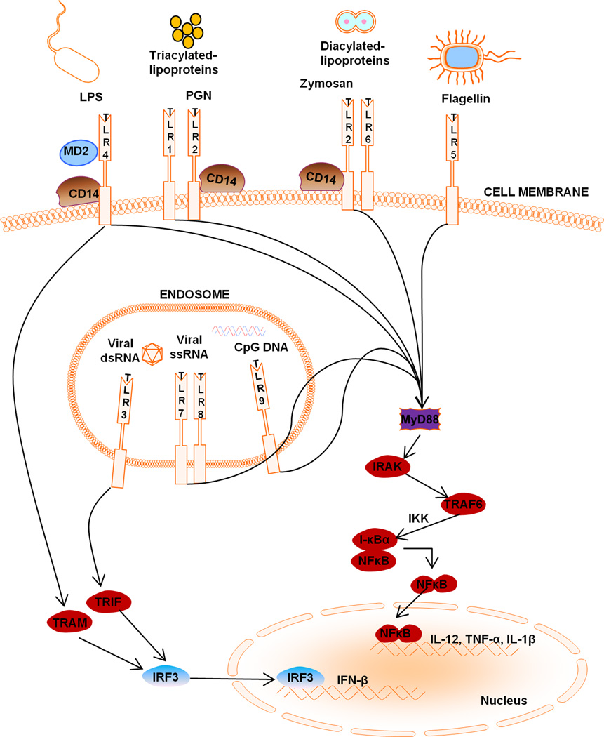
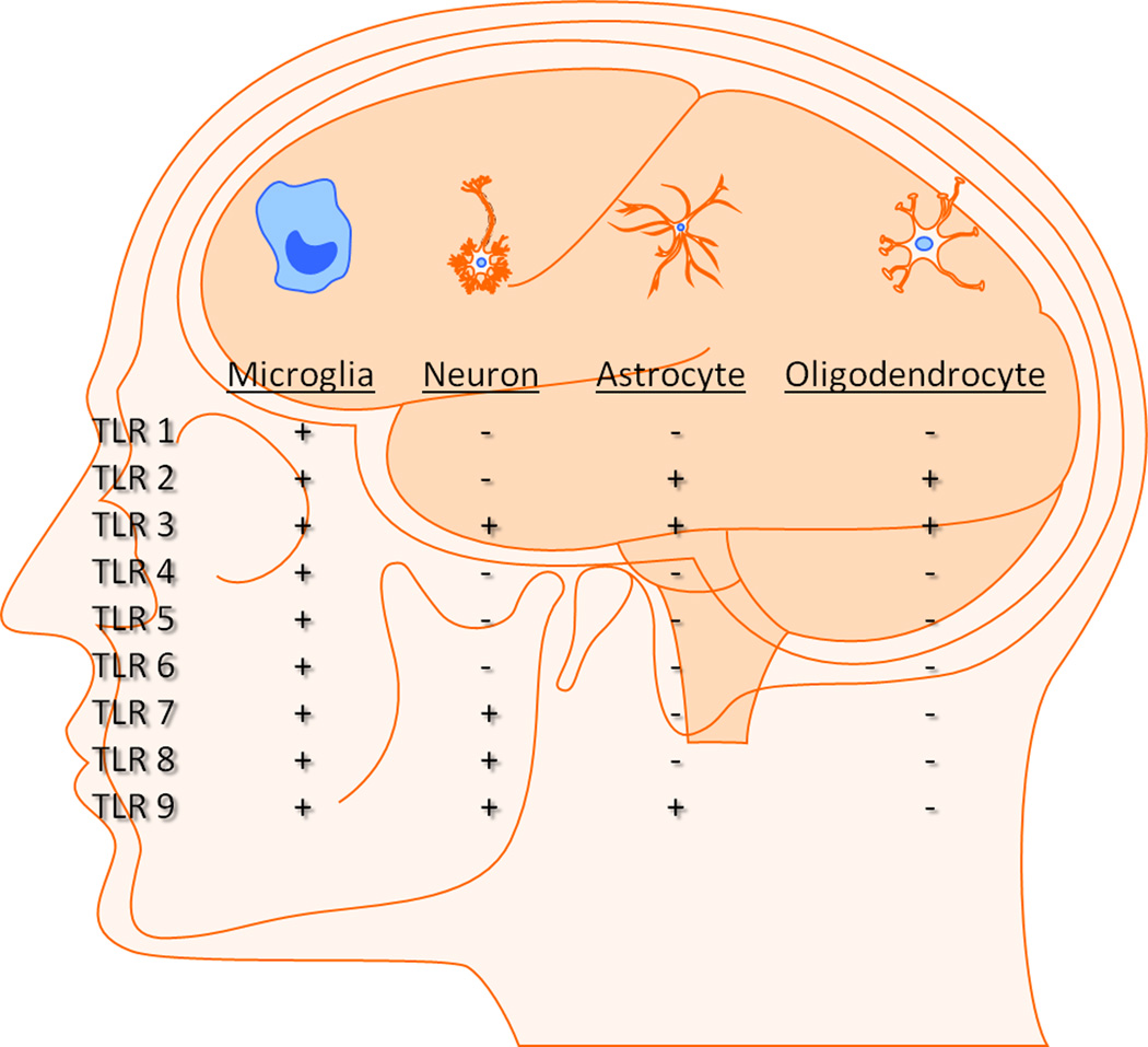
The discovery of mammalian TLRs (Toll-like receptors), first identified in 1997 based on their homology with Drosophila Toll, greatly altered our understanding of how the innate immune system recognizes and responds to diverse microbial pathogens. TLRs are evolutionarily conserved type I transmembrane proteins expressed in both immune and non-immune cells, and are typified by N-terminal leucine-rich repeats and a highly conserved C-terminal domain termed the TIR [Toll/interleukin (IL)-1 receptor] domain. Upon stimulation with their cognate ligands, TLR signalling elicits the production of cytokines, enzymes and other inflammatory mediators that can have an impact on several aspects of CNS (central nervous system) homoeostasis and pathology. For example, TLR signalling plays a crucial role in initiating host defence responses during CNS microbial infection. Furthermore, TLRs are targets for many adjuvants which help shape pathogen-specific adaptive immune responses in addition to triggering innate immunity. Our knowledge of TLR expression and function in the CNS has greatly expanded over the last decade, with new data revealing that TLRs also have an impact on non-infectious CNS diseases/injury. In particular, TLRs recognize a number of endogenous molecules liberated from damaged tissues and, as such, influence inflammatory responses during tissue injury and autoimmunity. In addition, recent studies have implicated TLR involvement during neurogenesis, and learning and memory in the absence of any underlying infectious aetiology. Owing to their presence and immune-regulatory role within the brain, TLRs represent an attractive therapeutic target for numerous CNS disorders and infectious diseases. However, it is clear that TLRs can exert either beneficial or detrimental effects in the CNS, which probably depend on the context of tissue homoeostasis or pathology. Therefore any potential therapeutic manipulation of TLRs will require an understanding of the signals governing specific CNS disorders to achieve tailored therapy.
Figures
References
-
- Medzhitov R, Janeway C. Innate immune recognition: mechanisms and pathways. Immunol. Rev. 2000;173:89–97. - PubMed
-
- Takeuchi O, Akira S. Pattern recognition receptors and inflammation. Cell. 2010;140:805–820. - PubMed
-
- Kaisho T, Akira S. Pleiotropic function of Toll-like receptors. Microbes Infect. 2004;6:1388–1394. - PubMed
-
- Qureshi ST, Medzhitov R. Toll-like receptors and their role in experimental models of microbial infection. Genes Immun. 2003;4:87–94. - PubMed
-
- Hoebe K, Beutler B. LPS, dsRNA and the interferon bridge to adaptive immune responses: Trif, Tram, and other TIR adaptor proteins. J. Endotoxin Res. 2004;10:130–136. - PubMed
Publication types
MeSH terms
Substances
Grants and funding
LinkOut - more resources
Full Text Sources
Other Literature Sources
Medical