. 2011 Jul 11:12:65.
doi: 10.1186/1471-2202-12-65.

Myosin VI contributes to synaptic transmission and development at the Drosophila neuromuscular junction

Affiliations

Myosin VI contributes to synaptic transmission and development at the Drosophila neuromuscular junction

Marta Kisiel et al. BMC Neurosci. .

Abstract

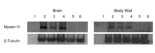
Background: Myosin VI, encoded by jaguar (jar) in Drosophila melanogaster, is a unique member of the myosin superfamily of actin-based motor proteins. Myosin VI is the only myosin known to move towards the minus or pointed ends of actin filaments. Although Myosin VI has been implicated in numerous cellular processes as both an anchor and a transporter, little is known about the role of Myosin VI in the nervous system. We previously recovered jar in a screen for genes that modify neuromuscular junction (NMJ) development and here we report on the genetic analysis of Myosin VI in synaptic development and function using loss of function jar alleles.

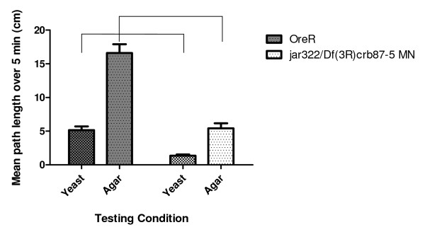
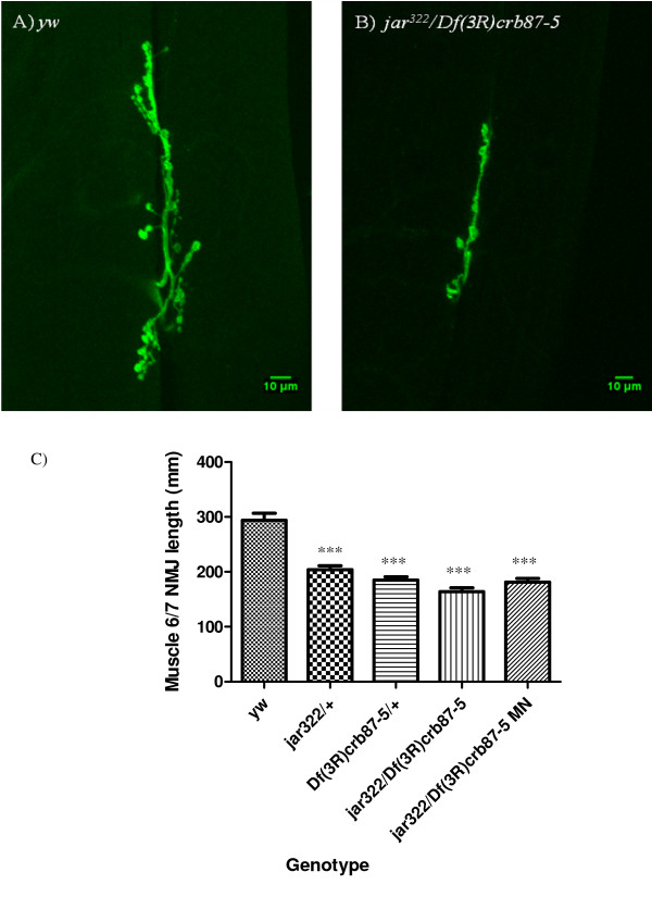
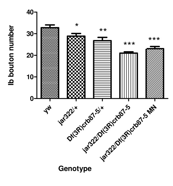
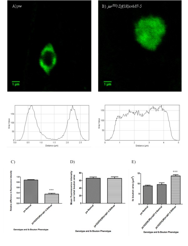
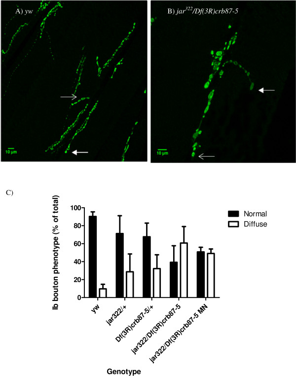
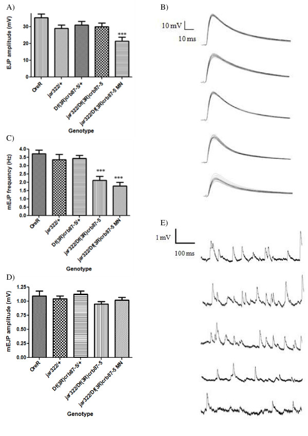
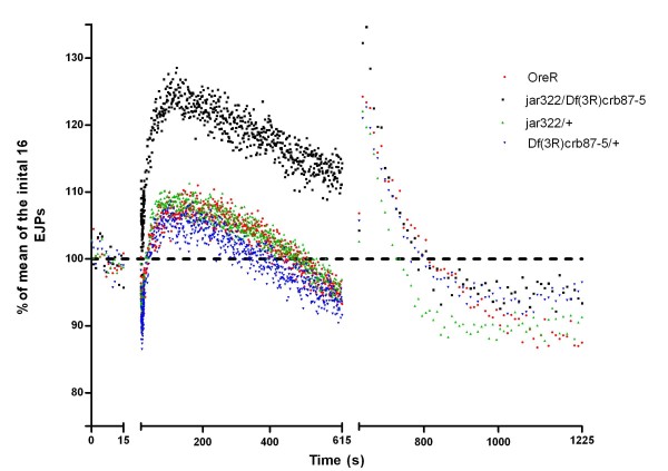
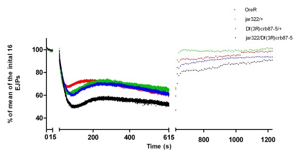
Results: Our experiments on Drosophila third instar larvae revealed decreased locomotor activity, a decrease in NMJ length, a reduction in synaptic bouton number, and altered synaptic vesicle localization in jar mutants. Furthermore, our studies of synaptic transmission revealed alterations in both basal synaptic transmission and short-term plasticity at the jar mutant neuromuscular synapse.

Conclusions: Altogether these findings indicate that Myosin VI is important for proper synaptic function and morphology. Myosin VI may be functioning as an anchor to tether vesicles to the bouton periphery and, thereby, participating in the regulation of synaptic vesicle mobilization during synaptic transmission.


Figures

Figure 1
Figure 1
Quantification of loss of function jaguar alleles. Western blotting of third instar larval brains and body walls revealed a reduction in Myosin VI levels in jar loss of function mutants. Lanes for both blots are numbered as follows: 1-ladder, 2-OreR, 3- jar322/+, 4- Df(3R)crb87-5/+, 5- jar322/Df(3R)crb87-5 and 6- jar322/Df(3R)crb87-5 MN. The loading control used was β-Tubulin. Relative Myosin VI proteins levels were first normalized to the level of β-tubulin and then quantified. Myosin VI levels were reduced in jar322/+ and Df(3R)crb87-5/+ larvae compared to the control in blots of both brain and body wall samples. No bands were observed for either brain or body wall samples for jar322/Df(3R)crb87-5 and jar322/Df(3R)crb87-5 MN larvae.
Figure 2
Figure 2
jaguar mutants exhibit general larval locomotor defects. Third instar jar322/Df(3R)crb87-5 MN larvae exhibited a significant decrease in path length on both a nutritive and non-nutritive substrate compared to the control (ANOVA, *** = p < 0.0001).
Figure 3
Figure 3
jaguar loss of function mutants display reduced muscle 6/7 NMJ length. Representative NMJs on ventral longitudinal muscles 6 and 7 from third instar Drosophila larvae of the control (A) and the jar loss of function mutant, jar322/Df(3R)crb87-5 (B). All images were acquired at the same magnification using a LSM510 confocal laser microscope. Muscle 6/7 NMJ lengths for the jar loss of function genotypes studied were significantly shorter than the control (Dunn's Multiple Comparison Test, *** = p < 0.001) (C). Bars represent mean ± SEM.
Figure 4
Figure 4
jaguar loss of function mutants exhibited a reduction in bouton number. A significant decrease in Ib bouton number was observed for all jar loss of function mutants studied at muscle 6/7 NMJs in segments A3, A4 and A5 compared to the control (Dunnett's Multiple Comparison Test, * = p < 0.05, ** = p < 0.01, *** = p < 0.001) (D). Bars represent mean ± SEM.
Figure 5
Figure 5
Synaptic vesicles are mislocalized at jaguar mutant boutons as revealed by immunostaining against synaptotagmin. A representative control Ib bouton and mutant jar322/Df(3R)crb87-5 Ib bouton are shown with their corresponding plot profiles (A and B). There was a significant reduction in relative difference in fluorescence intensity across jar322/Df(3R)crb87-5 mutant boutons (n = 13) compared to the control boutons (n = 25; Unpaired T-test, *** = p < 0.001) (C). Mean fluorescence intensity did not differ significantly between the mutant (n = 48) and the control (n = 64; Unpaired T-test, p > 0.05) (D). Diffuse jar322/Df(3R)crb87-5 boutons (n = 53) were significantly larger than control boutons (n = 64) and jar322/Df(3R)crb87-5 boutons with a normal, doughnut-shaped distribution of synaptotagmin (n = 27, Dunn's Multiple Comparison Test, *** = p < 0.001) (E). Boutons used for this study were from muscle 6/7 NMJs in segments A3, A4 and A5. Bars represent mean ± SEM.
Figure 6
Figure 6
Boutons exhibiting vesicle mislocalization increased corresponding to the severity of jaguar loss of function. Immunostaining against synaptotagmin of NMJs on muscles 6/7 of control and jar322/Df(3R)crb87-5 third instar larvae revealed different proportions of boutons exhibiting normal and diffuse synaptotagmin staining (A and B). Images were taken at different magnifications to visualize the entire NMJ. Open arrows indicate the normal, doughnut-shaped pattern of synaptotagmin localization and closed arrows indicate the diffuse pattern of synaptotagmin localization. Scoring the synaptotagmin distribution phenotype revealed a reduction in the percentage of normal boutons and an increase in the percentage of diffuse boutons in jar loss of function mutants compared to the control (C). Number of boutons analyzed were yw n = 48, jar322/+ n = 38, Df(3R)crb87-5/+ n = 28, jar322/Df(3R)crb87-5 n = 58 and jar322/Df(3R)crb87-5 MN n = 18. Boutons used for this study were from muscle 6/7 NMJs in segments A3, A4 and A5. Bars represent mean ± SEM.
Figure 7
Figure 7
jaguar loss of function mutants exhibited defects in basal synaptic transmission. Electrophysiological recordings from muscles 6/7 of third instar larvae revealed EJP amplitude was significantly reduced in the most severe jar mutant, jar322/Df(3R)crb87-5 MN, compared to the control (Dunnett's Multiple Comparison Test, *** = p < 0.001) (A). Sample traces of EJP recordings taken at 1 Hz, shown in the same order as listed for the graphs (B). mEJP frequency was significantly lower for jar322/Df(3R)crb87-5 and jar322/Df(3R)crb87-5 MN larvae compared to the control larvae (Dunnett's Multiple Comparison Test, *** = p < 0.001) (C). There was no significant difference in mEJP amplitude between jar mutants and the control (ANOVA, p > 0.05) (D). Sample traces of electrophysiological recordings from muscles 6/7 over 2 seconds, shown in the same order as listed for the graphs (E). Bars represent mean ± SEM.
Figure 8
Figure 8
High frequency stimulation in 1 mM Ca2+ saline revealed enhanced potentiation in jar322/Df(3R)crb87-5 mutants. Data is shown as a percent of the average of the first 16 evoked junctional potentials (EJPs) recorded at 1 Hz. This baseline recording of 16 EJPs at 1 Hz was followed by a stimulation of 10 Hz recorded over 10 minutes and terminated with a 10 minute recording at 0.1 Hz. Following the first 25 seconds from the onset of 10 Hz stimulation, every tenth recording is shown. A significant enhancement in EJP amplitude was observed for jar322/Df(3R)crb87-5 larvae during the first three minutes of the high frequency protocol compared to the control (Dunnett's Multiple Comparison Test, p < 0.001).
Figure 9
Figure 9
High frequency stimulation in 10 mM Ca2+ saline revealed enhanced depression in jar322/Df(3R)crb87-5 mutants. Data is shown as a percent of the average of the first 16 evoked junctional potentials (EJPs) recorded at 1 Hz. This baseline recording of 16 EJPs at 1 Hz was followed by a stimulation of 10 Hz recorded over 10 minutes and terminated with a 10 minute recording at 0.1 Hz. Following the first 25 seconds from the onset of 10 Hz stimulation, every tenth recording is shown. The initial depression in EJP amplitude measured relative to EJP amplitude at the onset of high frequency stimulation was significantly greater for jar322/Df(3R)crb87-5 larvae than the control larvae (ANOVA, p < 0.01).

Similar articles

Cited by

References

    1. Nunes P, Haines N, Kuppuswamy V, Fleet DJ, Stewart BA. Synaptic vesicle mobility and presynaptic F-actin are disrupted in a N-ethylmaleimide-sensitive factor allele of Drosophila. Mol Biol Cell. 2006;17(11):4709–4719. doi: 10.1091/mbc.E06-03-0253. - DOI - PMC - PubMed
    1. Seabrooke S, Qiu X, Stewart B. Nonmuscle Myosin II helps regulate synaptic vesicle mobility at the Drosophila neuromuscular junction. BMC Neuroscience. 2010;11(1):37. doi: 10.1186/1471-2202-11-37. - DOI - PMC - PubMed
    1. Gaffield MA, Betz WJ. Synaptic vesicle mobility in mouse motor nerve terminals with and without synapsin. J Neurosci. 2007;27(50):13691–13700. doi: 10.1523/JNEUROSCI.3910-07.2007. - DOI - PMC - PubMed
    1. Gaffield MA, Rizzoli SO, Betz WJ. Mobility of Synaptic Vesicles in Different Pools in Resting and Stimulated Frog Motor Nerve Terminals. Neuron. 2006;51(3):317–325. doi: 10.1016/j.neuron.2006.06.031. - DOI - PubMed
    1. Jordan R, Lemke EA, Klingauf J. Visualization of synaptic vesicle movement in intact synaptic boutons using fluorescence fluctuation spectroscopy. Biophys J. 2005;89(3):2091–2102. doi: 10.1529/biophysj.105.061663. - DOI - PMC - PubMed

Publication types

MeSH terms

LinkOut - more resources