. 2011 Oct;1813(10):1836-44.
doi: 10.1016/j.bbamcr.2011.06.012. Epub 2011 Jul 1.

HIV-1 Tat binds to SH3 domains: cellular and viral outcome of Tat/Grb2 interaction

Affiliations

HIV-1 Tat binds to SH3 domains: cellular and viral outcome of Tat/Grb2 interaction

Slava Rom et al. Biochim Biophys Acta. 2011 Oct.

Abstract

The Src-homology 3 (SH3) domain is one of the most frequent protein recognition modules (PRMs), being represented in signal transduction pathways and in several pathologies such as cancer and AIDS. Grb2 (growth factor receptor-bound protein 2) is an adaptor protein that contains two SH3 domains and is involved in receptor tyrosine kinase (RTK) signal transduction pathways. The HIV-1 transactivator factor Tat is required for viral replication and it has been shown to bind directly or indirectly to several host proteins, deregulating their functions. In this study, we show interaction between the cellular factor Grb2 and the HIV-1 trans-activating protein Tat. The binding is mediated by the proline-rich sequence of Tat and the SH3 domain of Grb2. As the adaptor protein Grb2 participates in a wide variety of signaling pathways, we characterized at least one of the possible downstream effects of the Tat/Grb2 interaction on the well-known IGF-1R/Raf/MAPK cascade. We show that the binding of Tat to Grb2 impairs activation of the Raf/MAPK pathway, while potentiating the PKA/Raf inhibitory pathway. The Tat/Grb2 interaction affects also viral function by inhibiting the Tat-mediated transactivation of HIV-1 LTR and viral replication in infected primary microglia.


Figures

Figure 1
Figure 1. Tat protein contains two putative SH3 domain-binding sites
A) Amino acid sequence of Tat showing two possible SH3 binding sites (in bold). B) List of SH3 domain containing proteins predicted to bind TAT using SH3 hunter web server (http://cbm.bio.uniroma2.it/SH3-Hunter/). First column represents amino acid sequence of peptide and its location in the Tat protein. Second column gives names of domains of interacting proteins. The last three columns in the output define respectively the significance (score) and the reliability (sensitivity and precision) of the prediction.
Figure 2
Figure 2. Tat directly interacts with Grb2
A) Scheme of GST-Tat full-length and various mutants cloned into pGex-5x-1 plasmid for GST-fusion protein expression. B) Western blot to detect Grb2 pulled-down using wild type Tat101 or the various mutants. C+ represents whole cell lysate (15 μg). C) GST-Grb2 pull-down of Tat from cells transfected with Tat bearing a single SH3-binding domain mutated (P3/6A and P81/84A, respectively), the double mutation (P3/6/81/84A) or the wild type Tat (WT). D) Scheme of GST-Grb2 mutants. Grb2 full-length and three mutants ΔN, ΔC, ΔN/C were cloned into pGex-5x-1 plasmid for GST-fusion protein expression. E) Western blot to detect Grb2 interaction with Tat in cells transfected with Tat101. F) Immunoblot shows that Grb2 wt and Grb2-SH3-C can efficiently pull down Gab1, while recombinant Tat competes with Gab1 for the binding to Grb2 (Panel G).
Figure 3
Figure 3. Detection of Grb2 and HIV-1 Tat in HIV-Encephalitis
Immunohistochemistry analysis shows the presence of Grb2 in the cytoplasm of reactive astrocytes in areas of neuro-inflammation (Panel A). The HIV-1 transactivator protein Tat is detected in endothelial cells, perivascular macrophages and robustly in the cytoplasm of reactive astrocytes in areas of encephalitis (Panel B). In contrast, non-affected areas of the brain within the same section show no expression of Tat (Panel C). Double labeling corroborates the modest presence of Grb2 (Panel D, rhodamine), and abundant expression of Tat (Panel E, fluorescein), in the cytoplasm of astrocytes, where the two proteins co-localize (Panel F), as demonstrated by deconvolution imaging (Inserts). G) Immunohistochemistry analysis to detect p24 in HIVE. H, I, J) Immunolabeling shows the presence of Tat in reactive astrocytes also positive for p24. Original magnification for all panels is 600x and 1000x in the inserts.
Figure 4
Figure 4. Co-localization of Grb2 and Tat in LN229 cells
Representative images show Tat and Grb2 co-localization in LN229 cells transfected with pEYFP and pEYFP-Tat, respectively. Note the diffuse cytoplasmic immunolabeling of Grb2 in control pEYFP transfected cells (Panels B and C), and its nuclear traslocation when Tat is present (Panels E and F). Original magnifications 100X.
Figure 5
Figure 5. Inhibitory effect of Tat in IGF-IR-mediate ERK1/2 phosphorylation
Representative Western blot showing activation of MAP kinases Erk1/2 by IGF-1 (40 ng/ml) in pEYFP or pEYFP-Tat expressing cells at the time points of 0, 2 and 10 minutes (upper panel). Total ERKs indicates equal loading (middle panel). Expression of IGF-IR is not affected by Tat (lower panel).
Figure 6
Figure 6. Phosphorylation of S259 inhibitory site of c-Raf is increased in Tat-transfected Glioblastoma LN229 cells
A) Western blot analysis to detect phosphorylation levels of c-Raf S259. Fold ratio of time 0 (lane 1) accepted as 1. B) Phosphorylation levels of c-Raf S259 and Erk1/2 in Tat overexpressing LN229 cells transfected together with Grb2 full-length and three mutants ΔN, ΔC, ΔN/C were detected by Western blot. Total c-Raf indicates equal loading. Experiments were repeated at least 3 times; representative Western blot is shown in the figure.
Figure 7
Figure 7. Tat-induced PKA activation mediates c-Raf (S259) phosphorylation
A) Western blot to detect c-Raf S259 phosphorylation and activation of Erk1/2 in LN229 cells transfected with pEYFP or pEYFP-Tat, pretreated with specific inhibitors (LY294002 50 μM, H89 25 μM) for 30 min and stimulated with IGF-1 for the indicated times. B) The kinase activity of PKA was tested in LN229 cells transfected with pEYFP, pEYFP-Tat, pEYFP-Tat together with Grb2 or Grb2 alone and stimulated with IGF-1 for 0 and 10 minutes. Activity was assayed using commercially available kit. Asterisks indicate values statistically significant (p<0.05). Ns: not statistically significant.
Figure 8
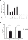
Figure 8. Suppression of LTR activity and inhibition of HIV-1 replication by overexpression of Grb2 protein
A) Promoter functional assay showing activation of HIV-LTR by Tat. LN229 cells were co-transfected with pEYFP-Tat, Grb2 wild type or truncated mutants, an HIV LTR–Firefly luciferase reporter plasmid and a Renilla luciferase control pRL-null plasmid. 24 hours later luciferase activity was measured according to manufacturer’s protocol. Inhibition of Tat-induced LTR by Grb2 and the indicated mutants is statistically significant: *p=0.00016, ** p=0.00005, # p=0.000316, ## p=0.00907. B) ELISA assay to detect p24 in the supernatant obtained from HIV-infected primary human fetal microglia collected at day 1 (D1) and day 3 (D3) post-infection with HIV. In the HIV/Grb2 sample cells were transfected with Grb2 full-length one day prior infection with HIV-1 JF-RL. Bar graph shows the average results from three independent experiments. Asterisk indicates p<0.001.

Similar articles

Cited by

References

    1. Mayer BJ. SH3 domains: complexity in moderation. J Cell Sci. 2001;114:1253–1263. - PubMed
    1. Pawson T. Protein modules and signalling networks. Nature. 1995;373:573–580. - PubMed
    1. Rickles RJ, Botfield MC, Zhou XM, Henry PA, Brugge JS, Zoller MJ. Phage display selection of ligand residues important for Src homology 3 domain binding specificity. Proc Natl Acad Sci U S A. 1995;92:10909–10913. - PMC - PubMed
    1. Sparks AB, Rider JE, Hoffman NG, Fowlkes DM, Quillam LA, Kay BK. Distinct ligand preferences of Src homology 3 domains from Src, Yes, Abl, Cortactin, p53bp2, PLCgamma, Crk, and Grb2. Proc Natl Acad Sci U S A. 1996;93:1540–1544. - PMC - PubMed
    1. Pawson T, Schlessingert J. SH2 and SH3 domains. Curr Biol. 1993;3:434–442. - PubMed

Publication types

MeSH terms

Substances