Review
. 2011 Aug 1;3(8):a003947.
doi: 10.1101/cshperspect.a003947.

Voltage-gated calcium channels

Affiliations
Review

Voltage-gated calcium channels

William A Catterall. Cold Spring Harb Perspect Biol. .

Abstract

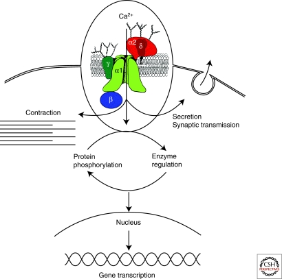
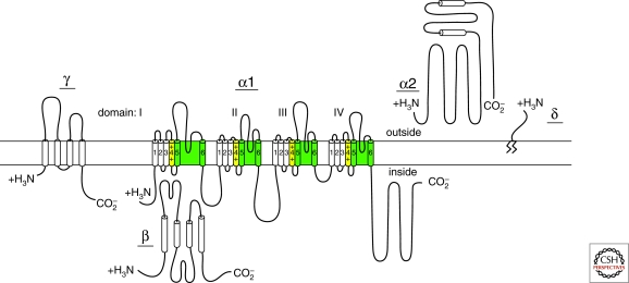
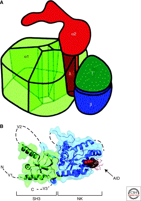
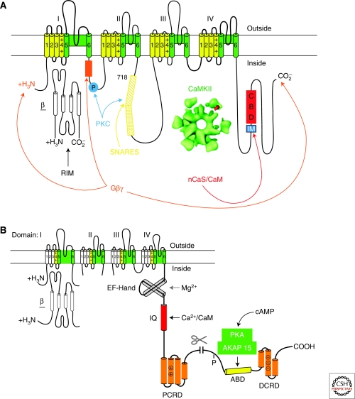
Voltage-gated calcium (Ca(2+)) channels are key transducers of membrane potential changes into intracellular Ca(2+) transients that initiate many physiological events. There are ten members of the voltage-gated Ca(2+) channel family in mammals, and they serve distinct roles in cellular signal transduction. The Ca(V)1 subfamily initiates contraction, secretion, regulation of gene expression, integration of synaptic input in neurons, and synaptic transmission at ribbon synapses in specialized sensory cells. The Ca(V)2 subfamily is primarily responsible for initiation of synaptic transmission at fast synapses. The Ca(V)3 subfamily is important for repetitive firing of action potentials in rhythmically firing cells such as cardiac myocytes and thalamic neurons. This article presents the molecular relationships and physiological functions of these Ca(2+) channel proteins and provides information on their molecular, genetic, physiological, and pharmacological properties.


Figures

Figure 1.
Figure 1.
Signal transduction by voltage-gated Ca2+ channels. Ca2+ entering cells initiates numerous intracellular events, including contraction, secretion, synaptic transmission, enzyme regulation, protein phosphorylation/dephosphorylation, and gene transcription. (Inset) Subunit structure of voltage-gated Ca2+ channels. The five-subunit complex that forms high-voltage-activated Ca2+ channels is illustrated with a central pore-forming α1 subunit, a disulfide-linked glycoprotein dimer of α2 and δ subunits, an intracellular β subunit, and a transmembrane glycoprotein γ subunit (in some Ca2+ channel subtypes). As described in the text, this model is updated from the original description of the subunit structure of skeletal muscle Ca2+ channels. (Adapted from Takahashi et al. 1987).
Figure 2.
Figure 2.
Subunit structure of Ca2+ channels. The structures of Ca2+ channel subunits are illustrated as transmembrane folding models; predicted α helices are depicted as cylinders; the lengths of lines correlate approximately to the lengths of the polypeptide segments represented; and the zigzag line on the δ subunit illustrates its glycophosphatidylinositol anchor.
Figure 3.
Figure 3.
Three-dimensional architecture of Ca2+ channels. (A) Illustration of the skeletal muscle CaV1.1 channel based on cryo-electronmicroscopy. This drawing assumes pseudo-fourfold symmetry of the α1 subunit. The view shows the extracellular side with the α2 subunit. The α1, γ, and δ subunits are embedded into the lipid membrane (not shown), which separates the extracellular α2 subunit from the cytosol. α2 is anchored via the disulfide-linked δ subunit within the α1 subunit. The proposed model allows for a tight interaction between α1 and δ as well as α1 and γ. (B) Structure of the CaVβ subunit with the α interaction domain (AID). Coordinates are for the CaVβ2a–CaV1.2 AID complex with SH3 (green) and NK (blue) domains are indicated. V1, V2, and V3 show the locations of the three variable domains that are absent from the structure. The AID (red) binds to a deep groove in the NK domain. AID residues tyrosine, tryptophan, and isoleucine are shown as CPK. The remaining residues are shown as lines.
Figure 4.
Figure 4.
Ca2+ channel signaling complexes. (A) The presynaptic Ca2+ channel signaling complex. A presynaptic Ca2+ channel α1 subunit is illustrated as a transmembrane folding diagram as in Figure 2. Sites of interaction of SNARE proteins (the synprint site), Gβγ subunits, protein kinase C (PKC), CaMKII, and CaM and CaS proteins are illustrated. IM, IQ-like motif; CBD, CaM binding domain. (B) The cardiac Ca2+ channel signaling complex. The carboxy-terminal domain of the cardiac Ca2+ channels is shown in expanded presentation to illustrate the regulatory interactions clearly. ABD, AKAP15 binding domain; DCRD, distal carboxy-terminal regulatory domain; PCRD, proximal carboxy-terminal regulatory domain; scissors, site of proteolytic processing. The DCRD binds to the PCRD through a modified leucine zipper interaction.

References

    1. Ahlijanian MK, Westenbroek RE, Catterall WA 1990. Subunit structure and localization of dihydropyridine-sensitive calcium channels in mammalian brain, spinal cord, and retina. Neuron 4: 819–832 - PubMed
    1. Arikkath J, Campbell KP 2003. Auxiliary subunits: Essential components of the voltage-gated calcium channel complex. Curr Opin Neurobiol 13: 298–307 - PubMed
    1. Armstrong CM, Bezanilla FM, Horowicz P 1972. Twitches in the presence of ethylene glycol bis (-aminoethyl ether)-N,N′-tetracetic acid. Biochim Biophys Acta 267: 605–608 - PubMed
    1. Artalejo CR, Adams ME, Fox AP 1994. Three types of calcium channel trigger secretion with different efficacies in chromaffin cells. Nature 367: 72–76 - PubMed
    1. Bajjalieh SM, Scheller RH 1995. The biochemistry of neurotransmitter secretion. J Biol Chem 270: 1971–1974 - PubMed

LinkOut - more resources