Review
. 2011 Jul 11;194(1):7-15.
doi: 10.1083/jcb.201102095.

Signal transduction by reactive oxygen species

Affiliations
Review

Signal transduction by reactive oxygen species

Toren Finkel. J Cell Biol. .

Abstract

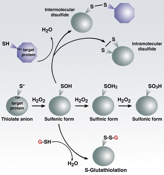
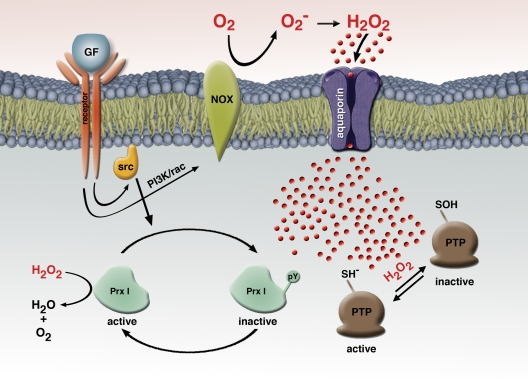
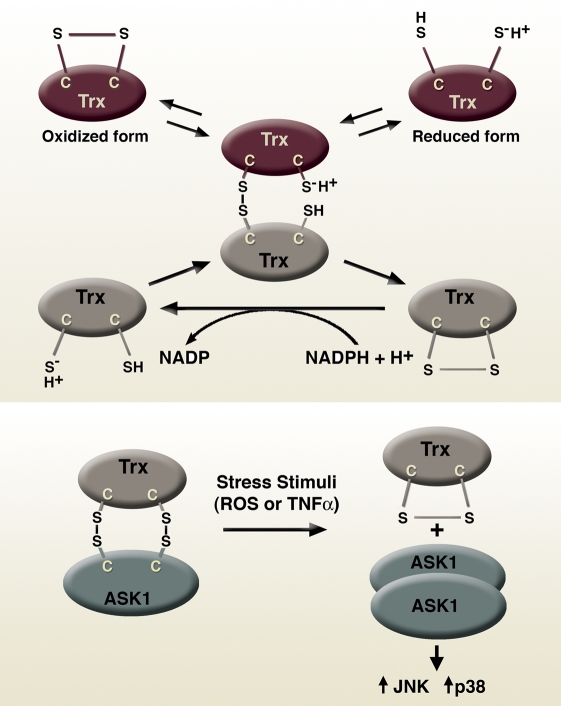
Although historically viewed as purely harmful, recent evidence suggests that reactive oxygen species (ROS) function as important physiological regulators of intracellular signaling pathways. The specific effects of ROS are modulated in large part through the covalent modification of specific cysteine residues found within redox-sensitive target proteins. Oxidation of these specific and reactive cysteine residues in turn can lead to the reversible modification of enzymatic activity. Emerging evidence suggests that ROS regulate diverse physiological parameters ranging from the response to growth factor stimulation to the generation of the inflammatory response, and that dysregulated ROS signaling may contribute to a host of human diseases.


Figures

Figure 1.
Figure 1.
Reactive oxygen species generation and disposal in the mitochondria. Primary sources of ROS occur from the transfer of electrons (e) to molecular oxygen at either Complex I or III. Superoxide produced at Complex I is thought to form only within the matrix, whereas at Complex III superoxide is released both into the matrix and the inner mitochondrial space (IMS). In addition to the cytochrome chain, ROS can be formed by enzymatic action of numerous enzymes including monoamine oxidase (MAO) and cytochrome b5 reductase (Cb5R) located on the outer mitochondrial membrane (OMM), as well as glycerol-3-phosphate dehydrogenase (GPDH) and in some cell types, various cytochrome P450 enzymes located in the inner mitochondrial membrane (IMM). There are also several matrix enzymes and complexes (box) including aconitase, pyruvate dehydrogenase (PDH), and α-ketoglutarate dehydrogenase (αKGDH) that can generate superoxide. Although one-electron reactions predominate, two-electron reactions leading to direct hydrogen peroxide production can occur as when, for instance, cytochrome c (Cyt C) and p66shc interact within the IMS. Once generated, superoxide is dismutated spontaneously or enzymatically by manganese superoxide dismutase (MnSOD). The hydrogen peroxide that is formed is further catabolized by the action of enzymes such as catalase (CAT), glutathione peroxidase (GPx), and peroxiredoxin 3 (Prx3). For further details see the text, as well as other recent reviews (Lin and Beal, 2006; Brand, 2010). CoQ, Coenzyme Q.
Figure 2.
Figure 2.
Cysteine biochemistry allows for redox-dependent signaling. Specific reactive cysteine (Cys) residues within target proteins can be covalently modified by oxidative stress. Much like phosphorylation on serine or threonine residues, alteration of the thiol group can in turn modify enzymatic activity. Although the sulfenic form (SOH) is readily reversible, higher states of oxidation generally, but not always, lead to irreversible modification.
Figure 3.
Figure 3.
Localization of oxidant signaling. One unresolved issue in redox signaling is how specificity is achieved with highly diffusible agents such as hydrogen peroxide. One mechanism may be to control the accumulation of ROS to discrete areas. Shown here is the signaling pathway envisioned to result after growth factor (GF) stimulation. Ligand binding to its receptor stimulates, through a PI3K/Rac-dependent process, superoxide production from the Nox family of NADPH-dependent oxidases. Extracellular oxidants are channeled back into the cell through specific plasma membrane aquaporins. Although hydrogen peroxide (red circles) can be rapidly and efficiently degraded by intracellular antioxidants, Src family members stimulated by growth factors appear to phosphorylate and subsequently inactivate the main intracellular peroxide scavenger, peroxiredoxin (Prx I). This Prx1 inactivation only occurs in the region surrounding the stimulated growth factor, thus allowing for the local accumulation of hydrogen peroxide. When the ROS reach sufficient levels, target molecules such as protein phosphatases (PTP) can be reversibly oxidized.
Figure 4.
Figure 4.
Antioxidant proteins can regulate signaling pathways. Proteins like thioredoxin (Trx) can function in cells to maintain redox balance by catalyzing the reduction of oxidized proteins (top). Trx can in turn be reduced through a process involving thioredoxin reductase and NADPH. In addition, Trx can also participate in redox signaling by directly binding to signaling intermediates such as ASK1 (bottom). In this case, the Trx1–ASK1 interaction is redox dependent and in turn modulates the capacity of ASK1 to activate downstream effectors such as p38 MAPK and the c-Jun N-terminal kinase (JNK).

Similar articles

Cited by

References

    1. Adler V., Yin Z., Fuchs S.Y., Benezra M., Rosario L., Tew K.D., Pincus M.R., Sardana M., Henderson C.J., Wolf C.R., et al. 1999. Regulation of JNK signaling by GSTp. EMBO J. 18:1321–1334 10.1093/emboj/18.5.1321 - DOI - PMC - PubMed
    1. Ago T., Liu T., Zhai P., Chen W., Li H., Molkentin J.D., Vatner S.F., Sadoshima J. 2008. A redox-dependent pathway for regulating class II HDACs and cardiac hypertrophy. Cell. 133:978–993 10.1016/j.cell.2008.04.041 - DOI - PubMed
    1. Aguirre J., Lambeth J.D. 2010. Nox enzymes from fungus to fly to fish and what they tell us about Nox function in mammals. Free Radic. Biol. Med. 49:1342–1353 10.1016/j.freeradbiomed.2010.07.027 - DOI - PMC - PubMed
    1. Alexander A., Cai S.L., Kim J., Nanez A., Sahin M., MacLean K.H., Inoki K., Guan K.L., Shen J., Person M.D., et al. 2010. ATM signals to TSC2 in the cytoplasm to regulate mTORC1 in response to ROS. Proc. Natl. Acad. Sci. USA. 107:4153–4158 10.1073/pnas.0913860107 - DOI - PMC - PubMed
    1. Anderson E.J., Lustig M.E., Boyle K.E., Woodlief T.L., Kane D.A., Lin C.T., Price J.W., III, Kang L., Rabinovitch P.S., Szeto H.H., et al. 2009. Mitochondrial H2O2 emission and cellular redox state link excess fat intake to insulin resistance in both rodents and humans. J. Clin. Invest. 119:573–581 10.1172/JCI37048 - DOI - PMC - PubMed

Publication types

Substances