. 2011 Oct;25(10):3496-504.
doi: 10.1096/fj.10-176305. Epub 2011 Jul 11.

Chondrogenesis by chemotactic homing of synovium, bone marrow, and adipose stem cells in vitro

Affiliations

Chondrogenesis by chemotactic homing of synovium, bone marrow, and adipose stem cells in vitro

Avital Mendelson et al. FASEB J. 2011 Oct.

Abstract

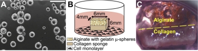
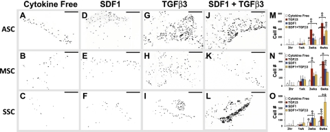
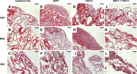
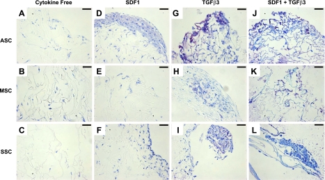
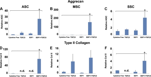
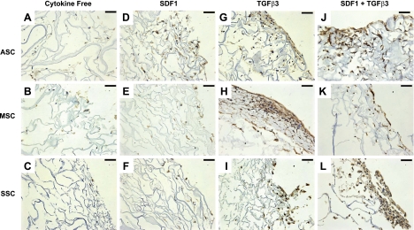
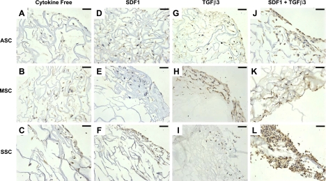
Cell transplantation has been well explored for cartilage regeneration. We recently showed that the entire articular surface of a synovial joint can regenerate by endogenous cell homing and without cell transplantation. However, the sources of endogenous cells that regenerate articular cartilage remain elusive. Here, we studied whether cytokines not only chemotactically recruit adipose stem cells (ASCs), mesenchymal stem cells (MSCs), and synovium stem cells (SSCs) but also induce chondrogenesis of the recruited cells. Recombinant human transforming growth factor-β3 (TGF-β3; 100 ng) and/or recombinant human stromal derived factor-1β (SDF-1β; 100 ng) was control released into an acellular collagen sponge cube with underlying ASCs, MSCs, or SSCs in monolayer culture. Although all cell types randomly migrated into the acellular collagen sponge cube, TGF-β3 and/or SDF-1β recruited significantly more cells than the cytokine-free control group. In 6 wk, TGF-β3 alone recruited substantial numbers of ASCs (558±65) and MSCs (302±52), whereas codelivery of TGF-β3 and SDF-1β was particularly chemotactic to SSCs (400±120). Proliferation of the recruited cells accounted for some, but far from all, of the observed cellularity. TGF-β3 and SDF-1β codelivery induced significantly higher aggrecan gene expression than the cytokine-free group for ASCs, MSCs, and SSCs. Type II collagen gene expression was also significantly higher for ASCs and SSCs by SDF-1 and TGF-β3 codelivery. Remarkably, the expression of aggrecan and type II collagen was detected among all cell types. Thus, homing of multiple stem/progenitor cell populations may potentially serve as an alternative or adjunctive approach to cell transplantation for cartilage regeneration.


Figures

Figure 1.
Figure 1.
Experimental schematics and tissue morphology. A) Light microscopy of microspheres with an average diameter of 70.8 ± 21.5 μm. Scale bar = 200 μm. B) Bilayered scaffold with an alginate layer containing gelatin microspheres overlaying a collagen sponge cube. Overall scaffold dimensions were 6 × 6 × 4 mm. Scaffolds were placed in the culture of ASCs, MSCs, and SSCs. C) Gross scaffold morphology following 3 wk culture. Glistening white tissue can be seen in the scaffold's collagen region.
Figure 2.
Figure 2.
Chemokine-mediated recruitment of ASCs, MSCs, and SSCs. After 6 wk in the culture of ASCs, MSCs, and SSCs, scaffolds were sectioned and stained with DAPI to visualize the cell nuclei. Sections from the top, middle, and bottom of the scaffold were stained (n=8–12). Four conditions were tested: cytokine free (A–C), SDF-1β (D–F), TGF-β3 (G–I), and TGF-β3 + SDF-1β (J–L) for ASCs (A, D, G, J), MSCs (B, E, H, K), and SSCs (C, F, I, L). All sections were quantified to determine the chemotactic effects of SDF-1β and/or TGF-β3 on ASCs (M), MSCs (N), and SSCs (O). Scale bars = 200 μm. Values shown are means ± se. Diamonds indicate significant difference over other conditions at same time point (P<0.05); circles indicate significant difference over previous time point (P<0.05).
Figure 3.
Figure 3.
Tissue formation on cytokine-induced cell recruitment. After 6 wk, scaffolds were stained with H&E for each of the 4 conditions tested: cytokine free (A–C), SDF-1β (D–F), TGF-β3 (G–I), and TGF-β3+SDF-1 (J–L) for ASCs (A, D, G, J), MSCs (B, E, H, K), and SSCs (C, F, I, L). Significant matrix deposition can be seen in ASCs due to SDF-1β (D), MSCs due to TGF-β3 (H), and SSCs due to SDF-1 + TGF-β3 (L), indicating that the scaffold substrate is conducive to cell attachment. Scale bars 50 μm.
Figure 4.
Figure 4.
Chondrogenic differentiation of chemokine-recruited cells. After 6 wk, sections were stained with toluidine blue for each of the 4 conditions tested: cytokine free (A–C), SDF-1β (D–F), TGF-β3 (G–I), and TGF-β3 + SDF-1β (J–L) for ASCs (A, D, G, J), MSCs (B, E, H, K), and SSCs (C, F, I, L). Scale bars = 50 μm. Positive staining reveals chondrogenesis among the TGF-β3-only group (G–I) and the SDF-1β + TGF-β3 (J–L) group for all 3 cell types.
Figure 5.
Figure 5.
Gene expression of aggrecan and type II collagen. After 6 wk, scaffolds were digested, and encapsulated cells were tested for aggrecan expression (A–C) and type II collagen (Col II; D–F) using RT-PCR (n=3). Relative aggrecan expression was significantly higher on TGF-β3 + SDF-1β codelivery for ASCs (A), MSCs (B), and SSCs (C) than the cytokine-free groups. TGF-β3 + SDF-1β codelivery also induced significantly higher Col II expression for ASCs (D) and SSCs (F) than the cytokine-free group. n.d., not detected (expression below detection limit).
Figure 6.
Figure 6.
Immunohistochemistry staining of aggrecan expression. After 6 wk, scaffold samples were immunostained with aggrecan antibody for each of the 4 conditions tested: cytokine free (A–C), SDF-1β (D–F), TGF-β3 (G–I), and TGF-β3 + SDF-1β (J–L) for ASCs (A, D, G, J), MSCs (B, E, H, K), and SSCs (C, F, I, L). Positive aggrecan staining revealed chondrogenesis among the TGF-β3-only group (G–I) and the SDF-1β + TGF-β3 group (J–L) for all 3 cell types. Scale bars = 50 μm.
Figure 7.
Figure 7.
Immunohistochemistry staining of type II collagen. After 6 wk, scaffold samples were immunostained with Col II for each of the 4 conditions: cytokine free (A–C), SDF-1β (D–F), TGF-β3 (G–I), and TGF-β3 + SDF-1β (J–L) for ASCs (A, D, G, J), MSCs (B, E, H, K), and SSCs (C, F, I, L). Positive Col II staining revealed chondrogenesis among the TGF-β3-only group for MSCs (H) and the SDF-1β + TGF-β3 group (J–L) for all 3 cell types. Scale bars = 50 μm.

Similar articles

Cited by

References

    1. Mao J. J. (2005) Stem-cell-driven regeneration of synovial joints. Biol. Cell 97, 289–301 - PubMed
    1. Dowthwaite G. P., Bishop J. C., Redman S. N., Khan I. M., Rooney P., Evans D. J., Haughton L., Bayram Z., Boyer S., Thomson B., Wolfe M. S., Archer C. W. (2004) The surface of articular cartilage contains a progenitor cell population. J. Cell Sci. 117, 889–897 - PubMed
    1. Raghunath J., Salacinski H. J., Sales K. M., Butler P. E., Seifalian A. M. (2005) Advancing cartilage tissue engineering: the application of stem cell technology. Curr. Opin. Biotechnol. 16, 503–509 - PubMed
    1. Chung C., Burdick J. A. (2008) Engineering cartilage tissue. Adv. Drug Deliv. Rev. 60, 243–262 - PMC - PubMed
    1. Fodor W. L. (2003) Tissue engineering and cell based therapies, from the bench to the clinic: the potential to replace, repair and regenerate. Reprod. Biol. Endocrinol. 1, 102 - PMC - PubMed

Publication types