. 2011 Sep;31(18):3773-89.
doi: 10.1128/MCB.05263-11. Epub 2011 Jul 11.

A novel role of vimentin filaments: binding and stabilization of collagen mRNAs

Affiliations

A novel role of vimentin filaments: binding and stabilization of collagen mRNAs

Azariyas A Challa et al. Mol Cell Biol. 2011 Sep.

Abstract

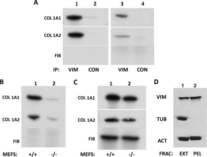
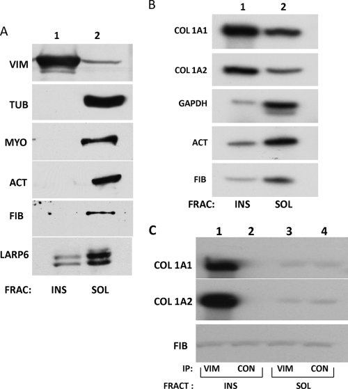
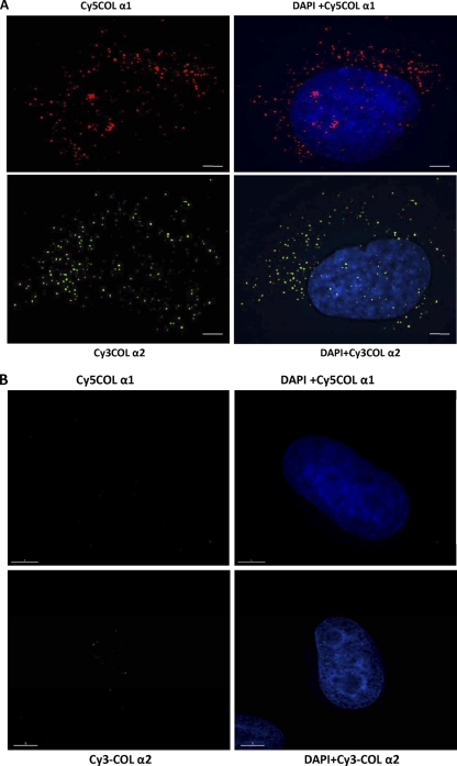
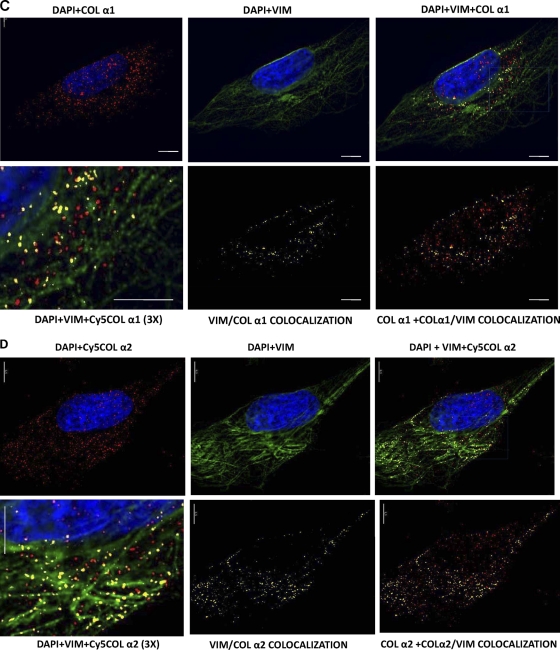
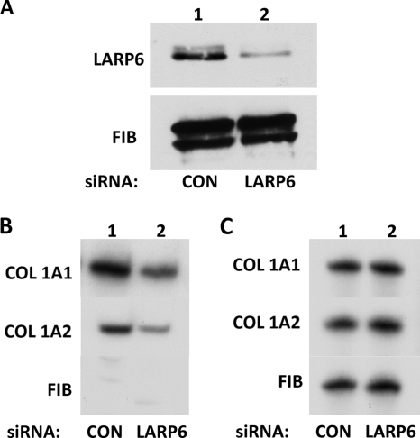
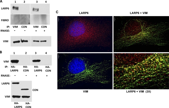
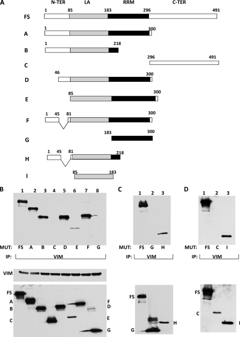
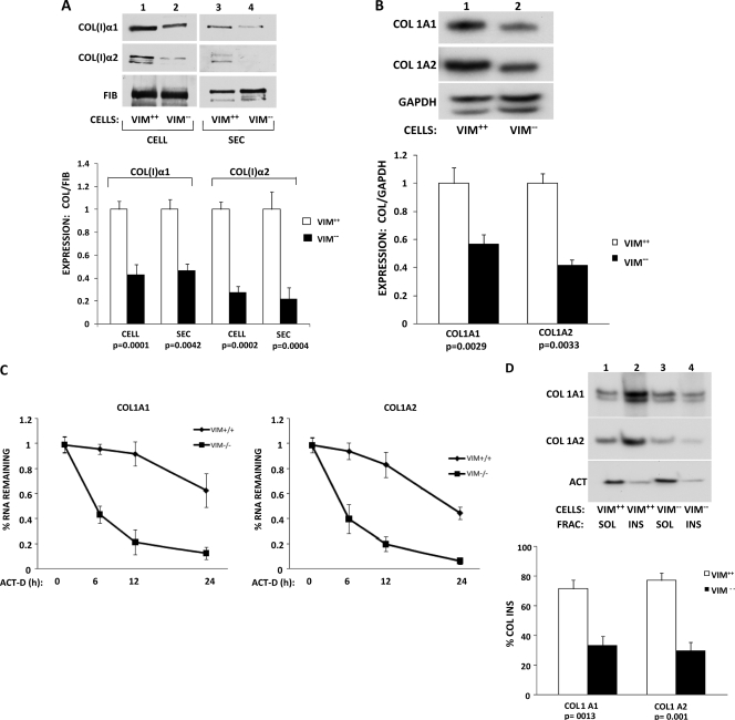
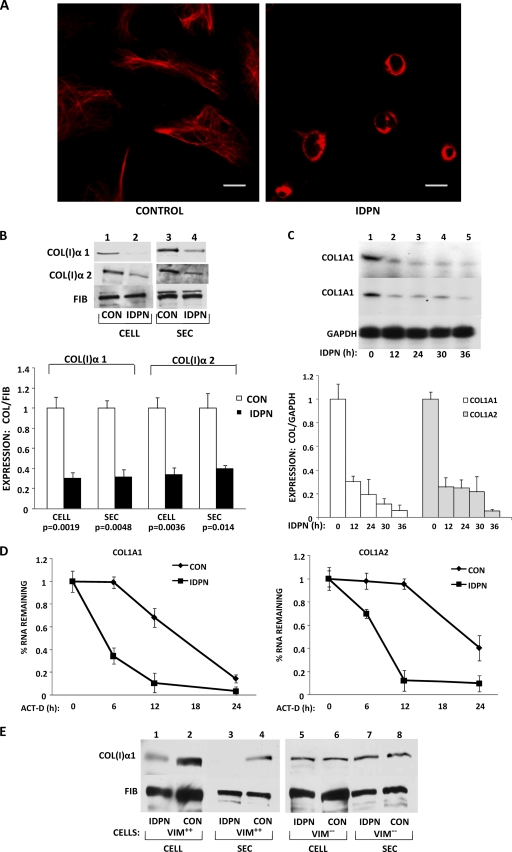
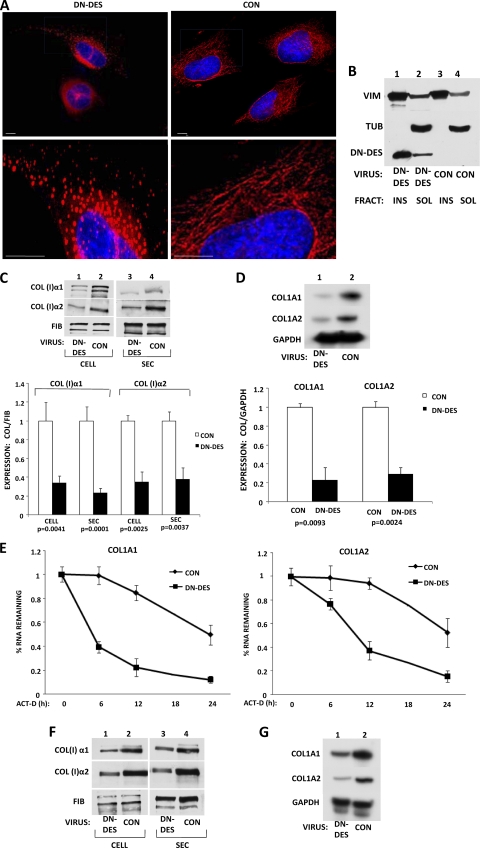
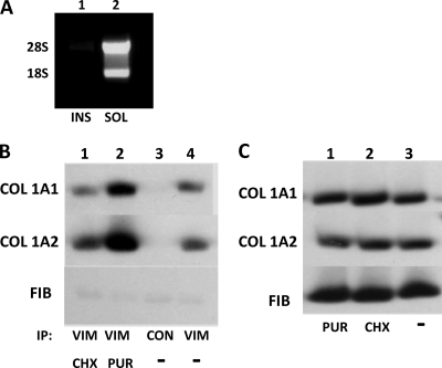
The stem-loop in the 5' untranslated region (UTR) of collagen α1(I) and α2(I) mRNAs (5'SL) is the key element regulating their stability and translation. Stabilization of collagen mRNAs is the predominant mechanism for high collagen expression in fibrosis. LARP6 binds the 5'SL of α1(I) and α2(I) mRNAs with high affinity. Here, we report that vimentin filaments associate with collagen mRNAs in a 5'SL- and LARP6-dependent manner and stabilize collagen mRNAs. LARP6 interacts with vimentin filaments through its La domain and colocalizes with the filaments in vivo. Knockdown of LARP6 by small interfering RNA (siRNA) or mutation of the 5'SL abrogates the interaction of collagen mRNAs with vimentin filaments. Vimentin knockout fibroblasts produce reduced amounts of type I collagen due to decreased stability of collagen α1(I) and α2(I) mRNAs. Disruption of vimentin filaments using a drug or by expression of dominant-negative desmin reduces type I collagen expression, primarily due to decreased stability of collagen mRNAs. RNA fluorescence in situ hybridization (FISH) experiments show that collagen α1(I) and α2(I) mRNAs are associated with vimentin filaments in vivo. Thus, vimentin filaments may play a role in the development of tissue fibrosis by stabilizing collagen mRNAs. This finding will serve as a rationale for targeting vimentin in the development of novel antifibrotic therapies.


Figures

Fig. 1.
Fig. 1.
Collagen α1(I) and collagen α2(I) mRNAs interact with vimentin in a 5′SL-dependent manner. (A) Pulldown of collagen mRNAs with an antivimentin antibody. Immunoprecipitation (IP) with an antivimentin antibody (VIM), followed by RT-PCR analysis of collagen (COL) α1(I), collagen α2(I), and fibronectin (FIB) mRNAs in human lung fibroblasts (lane 1) and scleroderma fibroblasts (lane 3). Lanes 2 and 4, control antibody (CON). Radiolabeled PCR products are indicated. (B) 5′SL-dependent interaction of collagen mRNAs with vimentin. The experiment was as in panel A, except extracts of wild-type (lane 1; +/+) and mutant (lane 2; −/−) mouse embryonic fibroblasts, which carry a mutation of the 5′ stem-loop in the collagen α1(I) gene, were used. (C) Total RNA of wild-type (lane 1; +/+) and mutant (lane 2; −/−) mouse embryonic fibroblasts analyzed by RT-PCR for collagen α1(I), collagen α2(I), and fibronectin mRNAs. (D) Presence of vimentin in the cell extract prepared by hypotonic and detergent lysis. The extract (EXT) and insoluble pellet (PEL) obtained after cell lysis were analyzed for vimentin, actin, and tubulin by Western blotting.
Fig. 2.
Fig. 2.
Collagen α1(I) and α2(I) mRNAs cofractionate with vimentin filaments. (A) Insoluble vimentin representing filaments (INS; lane 1) and soluble vimentin (SOL; lane 2) were fractionated by detergent lysis and centrifugation. The fractions (FRAC) were probed for the presence of vimentin (VIM), tubulin (TUB), myosin IIB (MYO), actin (ACT), fibronectin (FIB), and LARP6 proteins by Western blotting. (B) Collagen mRNAs segregate with insoluble vimentin. The fractions in panel A were analyzed by RT-PCR for collagen α1(I), collagen α2(I), GAPDH, actin, and fibronectin mRNAs. Shown is pulldown of collagen mRNAs with an antivimentin antibody. (C) Collagen mRNAs in the insoluble fraction coimmunoprecipitate with vimentin. The insoluble fraction (INS) was redissolved, and immunoprecipitation with vimentin antibody (lane 1) or control antibody (lane 2) was performed. (Lanes 3 and 4) The same experiment using the soluble fraction (SOL). The immunoprecipitated material was analyzed by RT-PCR for the presence of collagen α1(I), collagen α2(I), and fibronectin mRNAs.
Fig. 3.
Fig. 3.
Collagen mRNAs colocalize with vimentin intermediate filaments. (A) Visualization of collagen α1(I) mRNA (top) and collagen α2(I) mRNA (bottom) by RNA FISH. Collagen α1(I)-specific probes (labeled with Cy5; red) and collagen α2(I)-specific probes (labeled with Cy3; green) were hybridized to human lung fibroblasts. (B) The same experiment in HeLa cells, which do not express collagen α1(I) and α2(I) mRNAs. (C) Colocalization of collagen α1(I) mRNA with vimentin. RNA FISH for collagen α1(I) mRNA (upper left), immunostaining of vimentin (upper middle), and the overlaid image (upper right). Magnification (×3) of the selected area of the overlaid image is shown at the bottom left. The yellow dots indicate colocalization. (Bottom middle) Image showing only α1/VIM colocalization. (Bottom right) The image overlaid with total signal for α1(I) mRNA (upper left) to estimate the fraction of colocalized mRNA. Bars, 1 μm. (D) Colocalization of collagen α2(I) mRNA with vimentin. The same experiment as in panel C is shown, except collagen α2(I) mRNA was visualized.
Fig. 3.
Fig. 3.
Collagen mRNAs colocalize with vimentin intermediate filaments. (A) Visualization of collagen α1(I) mRNA (top) and collagen α2(I) mRNA (bottom) by RNA FISH. Collagen α1(I)-specific probes (labeled with Cy5; red) and collagen α2(I)-specific probes (labeled with Cy3; green) were hybridized to human lung fibroblasts. (B) The same experiment in HeLa cells, which do not express collagen α1(I) and α2(I) mRNAs. (C) Colocalization of collagen α1(I) mRNA with vimentin. RNA FISH for collagen α1(I) mRNA (upper left), immunostaining of vimentin (upper middle), and the overlaid image (upper right). Magnification (×3) of the selected area of the overlaid image is shown at the bottom left. The yellow dots indicate colocalization. (Bottom middle) Image showing only α1/VIM colocalization. (Bottom right) The image overlaid with total signal for α1(I) mRNA (upper left) to estimate the fraction of colocalized mRNA. Bars, 1 μm. (D) Colocalization of collagen α2(I) mRNA with vimentin. The same experiment as in panel C is shown, except collagen α2(I) mRNA was visualized.
Fig. 4.
Fig. 4.
LARP6-dependent association of collagen mRNAs with vimentin. (A) Knockdown of LARP6. Control siRNA (lane 1) and LARP6-specific siRNA (lane 2) were expressed in human lung fibroblasts, and the level of LARP6 was determined by Western blotting. Fibronectin was analyzed as a loading control. (B) LARP6 knockdown reduces the association of collagen mRNAs with vimentin. IP with an antivimentin antibody was performed in cells expressing control siRNA (lane 1) or LARP6-specific siRNA (lane 2). Collagen α1(I), collagen α2(I), and fibronectin were analyzed in the immunoprecipitate by RT-PCR. (C) Analysis of the input levels of mRNAs. Total mRNA before the IP was analyzed for expression of collagen α1(I), collagen α2(I), and fibronectin mRNAs by RT-PCR.
Fig. 5.
Fig. 5.
Interaction of LARP6 with vimentin. (A) Pulldown of endogenous LARP6 with anti-vimentin antibody. Shown is immunoprecipitation from lung fibroblasts with antivimentin antibody (lanes 1 and 3) or control antibody (lanes 2 and 4). Immunoprecipitates were left untreated (lanes 1 and 2) or treated with RNase A (lanes 3 and 4) prior to washing and Western blot analysis with anti-LARP6 antibody and antifibronectin (FIBRO) antibody. (Bottom) Input levels of vimentin. (B) Reverse experiment using HA-tagged LARP6. HA-tagged LARP6 (lanes 1 and 3) or control HA-tagged protein (lanes 2 and 4) was expressed in HEK293 cells, and IP was performed with anti-HA antibody without (lanes 1 and 2) or with (lanes 3 and 4) RNase A digestion. The immunoprecipitate was analyzed by Western blotting using antivimentin antibody. (Bottom) Input levels of transfected proteins and endogenous vimentin. (C) Colocalization of LARP6 with vimentin in lung fibroblasts. Immunostaining for vimentin (green; upper left) and endogenous LARP6 (red; upper right) and the merged image (lower left). The areas of colocalization are yellow. (Lower right) Higher magnification of the same image.
Fig. 6.
Fig. 6.
LARP6 interacts with vimentin through the La domain. (A) Schematic representation of the constructs used. FS, full-length LARP6. The domains of LARP6 are indicated, with amino acid numbering above. A to I, different deletion mutants of LARP6. All proteins had an HA tag at the N terminus (N-TER). C-TER, C terminus. (B) Interaction of FS LARP6 and deletion mutants with vimentin. (Top) FS LARP6 (lane 1) and the deletion mutants (lanes 2 to 8) were expressed in HEK293 cells. IP was done with antivimentin antibody and Western blotting with anti-HA antibody. (Bottom) Expression of the proteins in the input material. (C) Interaction of the extended La domain with vimentin. A construct containing the intact La domain with additional amino acids at the N terminus (H; lane 3) was analyzed for interaction with vimentin by IP. Lane 1, FS LARP6 as a positive control; lane 2, mutant G as a negative control. (Bottom) Expression of the proteins in the input material. (D) Interaction of the La domain only with vimentin. (Lane 1) Construct I, containing only the La domain, was analyzed for IP with vimentin. (Lane 2) Construct C as a negative control. (Bottom) Expression of the proteins in the input material.
Fig. 7.
Fig. 7.
Decreased collagen synthesis in vimentin knockout cells. (A) Expression of type I collagen in embryonic fibroblasts from wt (Vim+/+) and vimentin knockout (Vim−/−) mice. (Top) Representative Western blot of cellular (CELL; lanes 1 and 2) and secreted (SEC; lanes 3 and 4) levels of collagen (I) α1 and α2 polypeptides of Vim+/+ cells (lanes 1 and 3) and Vim−/− cells (lanes 2 and 4). Loading control, fibronectin. (Bottom) Quantitation of expression from three independent experiments. The expression of collagen polypeptides was normalized to the expression of fibronectin and arbitrarily set as 1 for Vim+/+ cells. The error bars represent SEM, and statistical significance is indicated. (B) Reduced steady-state levels of collagen α1(I) and α2(I) mRNAs in Vim−/− cells. (Top) Representative RT-PCR analysis of collagen α1(I), collagen α2(I), and GAPDH mRNA levels in Vim+/+ (lane 1) and Vim−/− (lane 2) mouse embryonic fibroblasts. (Bottom) Quantification of mRNA levels in three independent experiments. The expression of collagen mRNAs was normalized to the expression of GAPDH mRNA and arbitrarily set as 1 for Vim+/+ cells. The error bars represent SEM, and statistical significance is indicated. (C) Decay rates of collagen α1(I) and α2(I) mRNAs in Vim+/+ and Vim−/− cells. Transcription was blocked by actinomycin D (ACT-D) in Vim+/+ and Vim−/− cells, and collagen α1(I) and α2(I) mRNA levels were measured at 0 h, 6 h, 12 h, and 24 h after the block by RT-PCR. GAPDH mRNA was measured as a loading control. The expression of collagen mRNAs was normalized to the expression of GAPDH mRNA and set as 1 for Vim+/+ cells. The values for α1(I) mRNA (left) and α2(I) mRNA (right) were plotted for each time point, and error bars representing SEM are shown. (D) Segregation of collagen mRNAs with insoluble vimentin. (Top) Vimentin wt fibroblasts (Vim+/+; lanes 1 and 2) and vimentin knockout fibroblasts (Vim−/−; lanes 3 and 4) were fractionated into soluble (lanes 1 and 3) and insoluble (lanes 2 and 4) fractions. The fractions were analyzed for the presence of collagen α1(I), collagen α2(I), and actin mRNAs by RT-PCR. (Bottom) Percentages of collagen mRNAs found in the insoluble fractions of Vim+/+ and Vim−/− cells, as estimated by three independent experiments. Statistical significance and error bars representing SEM are shown.
Fig. 8.
Fig. 8.
Disruption of vimentin filaments by IDPN reduces collagen synthesis. (A) Collapse of vimentin filaments after IDPN treatment. Untreated human lung fibroblasts (HLF) (Control) and HLF treated with 1% IDPN were immunostained with antivimentin antibody. Bars, 1 μm. (B) IDPN reduces collagen protein. (Top) Cellular (lanes 1 and 2) and secreted (lanes 3 and 4) levels of collagen α1(I) and α2(I) polypeptides from control cells (lanes 1 and 3) or IDPN-treated cells (lanes 2 and 4) were analyzed by Western blotting. Loading control, fibronectin. (Bottom) Collagen expression was normalized to fibronectin expression and plotted from three independent experiments. The statistical significance and error bars representing SEM are shown. (C) IDPN decreases the steady-state levels of collagen mRNAs. (Top) Collagen mRNAs from control cells and cells treated with IDPN for the indicated times were analyzed by RT-PCR. Loading control, GAPDH. (Bottom) The expression of collagen mRNAs was normalized to the expression of GAPDH mRNA and plotted from three independent experiments. The error bars represent SEM. (D) Decay of collagen α1(I) mRNA (left) and collagen α2(I) mRNA (right) in control and IDPN-treated HLF. Transcription was blocked with actinomycin D, and the levels of collagen mRNAs were estimated by RT-PCR at the indicated times. The expression at time zero was set as 1. The error bars represent SEM estimated from three independent experiments. (E) IDPN affects collagen expression only in vimentin-expressing cells. Cellular and secreted levels of collagen α1(I) polypeptide were measured in VIM+/+ fibroblasts (lanes 1 to 4) and in VIM−/− fibroblasts (lanes 5 to 8) by Western blotting. The cells were treated with IDPN or left untreated, as indicated. Loading control, fibronectin.
Fig. 9.
Fig. 9.
Disruption of vimentin filaments by a dominant-negative mutant of desmin reduces collagen synthesis. (A) A dominant-negative mutant of desmin (DN-DES) disrupts vimentin filaments. HLF were transduced with control adenovirus (right) or adenovirus expressing DN-DES (left), and the cells were immunostained with antivimentin antibody. The lower images show higher magnification. (B) DN-DES cofractionates with insoluble vimentin. The cells in panel A were fractionated into vimentin-soluble and -insoluble fractions and analyzed by Western blotting using antivimentin, antitubulin, and anti-FLAG antibodies, which recognize DN-DES. (C) (Top) Collagen α1(I) and α2(I) polypeptides from HLF transduced with control (lanes 2 and 4) or DN-DES (lanes 1 and 3) adenovirus were analyzed by Western blotting. Loading control, fibronectin. (Bottom) Collagen expression was normalized to fibronectin expression and is shown for three independent experiments. The expression in control cells was set as 1, and statistical significance and error bars representing SEM are shown. (D) DN-DES decreases the steady-state levels of collagen mRNAs. (Top) The cells in panel C were analyzed for expression of collagen α1(I), collagen α2(I), and GAPDH mRNAs by RT-PCR. (Bottom) The expression of collagen mRNAs was normalized to the expression of GAPDH and plotted for three independent experiments. The expression in control cells was set as 1, and the statistical significance and error bars representing SEM are shown. (E) The levels of collagen α1(I) mRNA (left) and collagen α2(I) mRNA (right) were estimated in control and DN-DES-expressing cells after transcriptional blocking with actinomycin D for the indicated times. The expression at time zero was set as 1. The error bars represent SEM estimated from three independent experiments. (F) DN-DES decreases collagen expression in scleroderma fibroblasts. Cellular (lanes 1 and 2) and secreted (lanes 3 and 4) levels of collagen polypeptides of cells expressing DN-DES (lanes 1 and 3) or control protein (lanes 2 and 4) were analyzed by Western blotting. Fibronectin is shown as a loading control. (G) Reduced collagen mRNA levels in scleroderma fibroblasts expressing DN-DES. Total RNA from cells expressing DN-DES (lane 1) or control protein (lane 2) were analyzed by RT-PCR for collagen α1(I), collagen α2(I), and GAPDH mRNAs.
Fig. 10.
Fig. 10.
Vimentin associates with untranslated collagen mRNAs. (A) Absence of rRNA in the insoluble fraction containing filamentous vimentin. Soluble (lane 2) and insoluble (lane 1) fractions were prepared, and total RNA was extracted and analyzed by agarose gel electrophoresis and ethidium bromide staining. (B) Dissociation of polysomes increases the association of collagen mRNAs with vimentin. Shown is immunoprecipitation with antivimentin antibody (lanes 1, 2, and 4) or no antibody (lane 3) from human lung fibroblasts treated with cycloheximide (CHX) (lane 1) or puromycin (PUR) (lane 2) or from untreated cells (−) (lanes 3 and 4). The immunoprecipitated material was analyzed for collagen α1(I), collagen α2(I), and fibronectin mRNAs by RT-PCR. (C) Cycloheximide and puromycin do not change the total levels of collagen mRNAs. Shown is RT-PCR analysis of collagen α1(I), collagen α2(I), and fibronectin mRNAs in cells treated with puromycin (lane 1) or cycloheximide (lane 2) or untreated cells (lane 3).

References

    1. . 2000. The American Association for the Study of Liver Diseases (AASLD) 51st annual meeting and postgraduate courses. October 27-31, 2000, Dallas, Texas, U. S. A. Abstracts. Hepatology 32:163A–655A - PubMed
    1. Bassell G., Singer R. H. 1997. mRNA and cytoskeletal filaments. Curr. Opin. Cell Biol. 9:109–115 - PubMed
    1. Bassell G. J., Powers C. M., Taneja K. L., Singer R. H. 1994. Single mRNAs visualized by ultrastructural in situ hybridization are principally localized at actin filament intersections in fibroblasts. J. Cell Biol. 126:863–876 - PMC - PubMed
    1. Ben-Ari Y., et al. 2010. The life of an mRNA in space and time. J. Cell Sci. 123:1761–1774 - PMC - PubMed
    1. Bitterman P. B., Henke C. A. 1991. Fibroproliferative disorders. Chest 99:81S–84S - PubMed

Publication types

MeSH terms