. 2011 Oct;31(19):4036-51.
doi: 10.1128/MCB.01342-10. Epub 2011 Jul 11.

A pathway for the control of anoikis sensitivity by E-cadherin and epithelial-to-mesenchymal transition

Affiliations

A pathway for the control of anoikis sensitivity by E-cadherin and epithelial-to-mesenchymal transition

Sanjeev Kumar et al. Mol Cell Biol. 2011 Oct.

Abstract

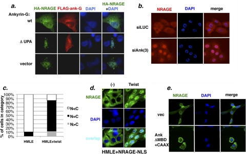
Detachment of epithelial cells from matrix or attachment to an inappropriate matrix engages an apoptotic response known as anoikis, which prevents metastasis. Cellular sensitivity to anoikis is compromised during the oncogenic epithelial-to-mesenchymal transition (EMT), through unknown mechanisms. We report here a pathway through which EMT confers anoikis resistance. NRAGE (neurotrophin receptor-interacting melanoma antigen) interacted with a component of the E-cadherin complex, ankyrin-G, maintaining NRAGE in the cytoplasm. Oncogenic EMT downregulated ankyrin-G, enhancing the nuclear localization of NRAGE. The oncogenic transcriptional repressor protein TBX2 interacted with NRAGE, repressing the tumor suppressor gene p14ARF. P14ARF sensitized cells to anoikis; conversely, the TBX2/NRAGE complex protected cells against anoikis by downregulating this gene. This represents a novel pathway for the regulation of anoikis by EMT and E-cadherin.


Figures

Fig. 1.
Fig. 1.
Ankyrin-G is downregulated by EMT and regulates anoikis sensitivity. (a) Ankyrin-G is downregulated by EMT. The indicated HMLE-derived cell lines were analyzed for ankyrin-G (∼210-kDa) protein by Western blotting or for ankyrin-G mRNA by RT-PCR. (b) Ankyrin-G promotes anoikis sensitivity: siRNA depletion in normal epithelial cells. HMLER cells transfected with two ankyrin-G siRNAs were assayed for anoikis. Western blotting (lower panel) confirmed the knockdown of ankyrin-G. (c) Ankyrin-G promotes anoikis sensitivity: ankyrin reexpression in EMT-derived cells. HMLE+shEcad cells or HMLE+shEcad+ankyrinΔMBD-CAAX cells were assayed for anoikis.
Fig. 2.
Fig. 2.
Ankyrin-G interacts with NRAGE. (a) Endogenous NRAGE–ankyrin-G interaction. Lysates of MCF10a cells or HMLE cells were immunoprecipitated with ankyrin-G pAb or nonimmune rabbit IgG (“con”) and probed for NRAGE. A no-lysate (NL) control was used to exclude IgG bands. WCL, whole-cell lysate representing 1/40 of input. (b) The MAGE and C-terminal domains of NRAGE interact with ankyrin-G. The indicated deletion mutants of NRAGE were cotransfected into 293T cells as GST fusion constructs, together with 3×HA-ankyrin p105. Western blots of glutathione-Sepharose-precipitates were analyzed for HA-ankyrin or GST-NRAGE, and each HA signal was normalized for its corresponding GST signal, producing the ratios shown in the graph. (c) The UPA domain of ankyrin-G interacts with NRAGE. The ankyrin-G domains indicated in the diagram, expressed as 3×HA-tagged proteins, were cotransfected with GST-NRAGE into 293T cells. Glutathione-Sepharose precipitates were probed for HA-ankyrin. The ratios of the HA signal in the precipitates versus the HA signal in the total lysates are shown. Note that the ankyrin repeat and ZU-5a domains were found to be dispensable for NRAGE interaction in an earlier experiment (data not shown).
Fig. 3.
Fig. 3.
Ankyrin-G sequesters NRAGE in the cytoplasm, and EMT enhances the accumulation of NRAGE in the nucleus. (a) Overexpressed ankyrin-G sequesters NRAGE in the cytoplasm. HT1080 cells were cotransfected with expression constructs encoding FLAG–tagged human ankyrin-G (210-kDa isoform), an NRAGE-nonbinding ankyrin-G deletion mutant, ΔUPA, or empty vector, together with HA-NRAGE. The cells were then fixed and stained for FLAG-ankyrin-G, HA-NRAGE, or chromatin (DAPI). (b) Depletion of ankyrin-G facilitates the accumulation of NRAGE in the nucleus. HMLE cells were transfected with multiple siRNAs for ankyrin-G (“si-Ank”) or luciferase (“si-Luc”), treated with leptomycin B, and stained for NRAGE (red) or chromatin (DAPI, blue). For the merged images, the color intensities were adjusted to provide optimal color balance. (c) EMT enhances the nuclear localization of NRAGE. HMLE or HMLE+Twist cells expressing HA-NRAGE (at ∼2-fold above the endogenous level for Twist) were treated with leptomycin B and stained for HA or chromatin. The histogram represents the frequency of cells with mostly nuclear staining (N>C), similar nuclear and cytoplasmic staining (N∼C), or mostly cytoplasmic staining (Nn = 88 for HMLE, n = 113 for Twist [P < 0.001]). (d) NRAGE-NLS is sequestered in the cytoplasm in normal cells and translocates to the nucleus in response to EMT. HMLE or HMLE+Twist cells stably transduced with an HA-NRAGE-NLS-expressing retrovirus were treated with leptomycin B and stained for NRAGE (green) and chromatin (DAPI, blue). (e) Ankyrin-G that is reexpressed in EMT-derived cells sequesters NRAGE in the cytoplasm. ΑnkyrinΔMBD+CAAX (see the text) was stably expressed in HMLE+shEcad cells, which were stained with NRAGE antibody.
Fig. 4.
Fig. 4.
NRAGE is an anoikis suppressor. (a) NRAGE depletion sensitizes HMLE+shEcad cells to anoikis. HMLE+shEcad cells were transfected with a pool of four siRNAs (upper panel) or a distinct additional siRNA (NRAGE-S2) and assayed for anoikis. (b) NRAGE depletion sensitizes HMLE+Twist cells to anoikis. HMLE+Twist cells were transfected with a pool of four siRNAs (upper panel) or infected with a doxycycline-inducible NRAGE shRNA lentivirus and grown for 3 days in the presence or absence of doxycycline (lower panel) and assayed for anoikis.
Fig. 5.
Fig. 5.
NRAGE is overexpressed in certain cancer types. (a) NRAGE mRNA is overexpressed in breast cancer. A panel of 5 normal and 43 breast cancer cDNAs was probed using TaqMan for NRAGE. β-Actin-normalized expression units are shown. (b) NRAGE mRNA is overexpressed in lung cancer. A panel of 8 normal and 40 lung cancer cDNAs was probed using TaqMan for NRAGE. β-Actin-normalized expression units are shown. (c) NRAGE protein is overexpressed in colon cancer. A tissue microarray containing 19 colon cancer samples and 6 normal samples was probed and quantitated by using AQUA (P < 0.006). (d) NRAGE protein is overexpressed in melanoma metastases. A tissue microarray containing samples of normal nevi (n = 8), primary melanoma (n = 9), or metastatic melanoma (n = 9) was probed and quantitated using AQUA (P < 0.009 for the difference in metastases versus nevi). (e) NRAGE protein is overexpressed in prostate cancer. A tissue microarray containing 17 prostate cancer samples and 6 normal samples was probed and quantitated by using AQUA (P < 0.08). (f). EMT upregulates NRAGE expression. HMLE cells or derivates expressing E-cadherin shRNA (“HMLE/shEcad”) or the Twist oncogene (“HMLE/Twist”) were probed for expression of NRAGE or, as a loading control, β-actin.
Fig. 6.
Fig. 6.
NRAGE interacts with TBX2, with functional consequences for anoikis sensitivity. (a). Endogenous interaction. HMLE+shEcad cells were immunoprecipitated with NRAGE pAb (“NR”) or an equal amount of control rabbit IgG (“Con”), and immunoprecipitates or total lysates were analyzed on a Western blot for TBX2 or NRAGE. (b) The C-terminal domain of NRAGE interacts with TBX2. The indicated deletion mutants (diagrammed in Fig. 2) of GST-NRAGE were cotransfected with FLAG-TBX2, and protein lysates were immunoprecipitated with FLAG antibody and analyzed on a Western blot for FLAG and GST. The ratios of GST to FLAG signal were quantitated and plotted to indicate the relative degree of interaction for each deletion construct, normalized against the expression level of each NRAGE deletion mutant (arbitrary units). (c) The repression domain of TBX2 interacts with NRAGE. 293T cells were cotransfected with expression constructs containing the indicated FLAG-tagged TBX2 constructs, and FLAG-agarose precipitates were analyzed for endogenous NRAGE protein by Western blotting. (d) NRAGE-TBX2 interaction mediates protection against anoikis. NRAGE−/− MEFs that were rescued with wild-type NRAGE (WT) or with the C-terminal deletion mutant that failed to interact with TBX2 (ΔC) were assayed for anoikis. (e) TBX2 is an anoikis suppressor. HMLE+shEcad cells transfected with TBX2 siRNA (Smartpool) or luciferase siRNA were assayed for anoikis at 8 h of suspension. Note that the efficiency of TBX2 knockdown (Western blot in the inset) correlated well with the magnitude of protection against anoikis.
Fig. 7.
Fig. 7.
NRAGE represses p14ARF expression in cells resulting from EMT and tumor cell lines. (a) HMLE cell lines expressing shEcad or Twist (upper panel), MDA-MB-435 breast carcinoma cells, or NCI-H1299 lung carcinoma cells (middle panel) were transfected with NRAGE Smartpool siRNA and analyzed for p14ARF protein induction. The normalized fold induction relative to the caspase-8 control is shown. For the lower panel, HMLE+Twist cells that expressed a doxycycline-inducible lentiviral NRAGE shRNA construct were assayed for p14ARF expression with or without doxycycline induction. (b) NRAGE cooperates with TBX2 to repress the p14ARF promoter. For the left panel, the Gal4-TBX2-repression domain (amino acids 515 to 574) was cotransfected at the indicated input DNA amounts with a Gal4-thymidine kinase-luciferase reporter into HMLE+shEcad cells that had been depleted of endogenous NRAGE with Smartpool siRNA or a nontargeting (NT) control siRNA. For the right panel, TBX2 was cotransfected at the indicated input DNA amounts with an ARF promoter-luciferase construct, with wild-type (first four pairs) or mutant TBX2 (fifth pair) binding sites, into 293T cells. The fold repression in cells pretransfected with control siRNA or NRAGE siRNA is shown at various input amounts of Gal4-TBX2-RD or TBX2 DNA. The enhancement of repression was calculated by dividing the fold repression due to Gal4-TBX2-RD in the control-siRNA samples by the fold repression due to Gal4-TBX2-RD in the NRAGE-siRNA samples. The results are averaged over two independent experiments. (c) TBX2 recruits NRAGE to the p14ARF promoter. For the left-hand panel, 293T cells were cotransfected with p14ARF-luciferase constructs containing the wild-type promoter (wt) or a mutant promoter (mut) lacking the previously characterized TBX2 binding site. FLAG-NRAGE and untagged TBX2 were cotransfected as indicated, and ChIP analysis was performed on FLAG-agarose precipitates. For the right-hand panel, a 3×Gal4-thymidine kinase promoter-luciferase construct was cotransfected with Gal4-TBX2 and FLAG-NRAGE, followed by ChIP analysis as described above. (d) EMT increases the occupancy of the p14ARF promoter by NRAGE. HMLE or HMLE+Twist cells that expressed similar levels of 3×FLAG-NRAGE (Western blot, right) were analyzed by ChIP using a FLAG antibody, a histone H3 antibody (as a positive control), or an HA antibody (as a negative control) and PCR primers spanning the TBX2 binding site of the p14ARF promoter.
Fig. 8.
Fig. 8.
p14ARF is downregulated by EMT and upregulated by ankyrin-G. (a) Downregulation by EMT. HMLE cells expressing shEcad (analyzed on one Western blot) or Twist (analyzed on another Western blot) were assayed for p14ARF protein (left panel) or mRNA (right panel). (b) Upregulation by ankyrin-G. HMLE+shEcad cells that express the ankyrinΔMBD-CAAX construct (see the text) were assayed for p14ARF protein expression. The normalized fold induction is shown.
Fig. 9.
Fig. 9.
p14ARF promotes anoikis sensitivity, and NRAGE confers anoikis resistance by suppressing p14ARF. (a) siRNA-mediated depletion of p14ARF confers anoikis resistance. HMLER cells transfected with a p14ARF exon 1β-specific siRNA that did not target p16 were assayed for anoikis at 24 h of suspension. Confirmation of the knockdown is shown in the lower panel. (b) ARF−/− and matched ARF+/+ MEFs were assayed for anoikis at 6 or 24 h of suspension. (c) NRAGE confers anoikis resistance by suppressing p14ARF (double-knockdown approach). HMLE+Twist cells that were depleted of NRAGE, p14ARF, or both were assayed for anoikis and p14ARF expression. (d) NRAGE confers anoikis resistance by suppressing p14ARF (complementation approach). HMLE+Twist-ER cells (induced to undergo EMT with 4-OHT) that expressed a doxycycline-inducible 6×HA-p14ARF construct were induced with doxycycline and assayed for anoikis compared against a control cell line with the empty vector. The Western blot shows that the induced level of 6×HA-p14ARF was similar to that expressed by HMLE-Twist cells prior to EMT induction.
Fig. 10.
Fig. 10.
Pathway leading from EMT to anoikis resistance.

Similar articles

Cited by

References

    1. Ayyanathan K., et al. 2007. The Ajuba LIM domain protein is a corepressor for SNAG domain mediated repression and participates in nucleocytoplasmic shuttling. Cancer Res. 67:9097–9106 - PubMed
    1. Barker P. A., Salehi A. 2002. The MAGE proteins: emerging roles in cell cycle progression, apoptosis, and neurogenetic disease. J. Neurosci. Res. 67:705–712 - PubMed
    1. Barrallo-Gimeno A., Nieto M. A. 2005. The Snail genes as inducers of cell movement and survival: implications in development and cancer. Development 132:3151–3161 - PubMed
    1. Bartel P. L., Fields S. 1995. Analyzing protein-protein interactions using two-hybrid system. Methods Enzymol. 254:241–263 - PubMed
    1. Bertrand M. J., et al. 2008. NRAGE, a p75NTR adaptor protein, is required for developmental apoptosis in vivo. Cell Death Differ. 15:1921–1929 - PMC - PubMed

Publication types

MeSH terms