

Tumor-associated macrophages regulate tumorigenicity and anticancer drug responses of cancer stem/initiating cells
- PMID: 21746895
- PMCID: PMC3145680
- DOI: 10.1073/pnas.1106645108
Tumor-associated macrophages regulate tumorigenicity and anticancer drug responses of cancer stem/initiating cells
Abstract
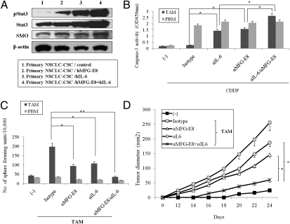
Recent evidence has unveiled the critical role of tumor cells with stem cell activities in tumorigenicity and drug resistance, but how tumor microenvironments regulate cancer stem/initiating cells (CSCs) remains unknown. We clarified the role of tumor-associated macrophages (TAMs) and their downstream factor milk-fat globule-epidermal growth factor-VIII (MFG-E8) in the regulation of CSC activities. Bone marrow chimeric systems and adoptive cell transfers elucidated the importance of MFG-E8 from TAMs in conferring to CSCs with the ability to promote tumorigenicity and anticancer drug resistance. MFG-E8 mainly activates signal transducer and activator of transcription-3 (Stat3) and Sonic Hedgehog pathways in CSCs and further amplifies their anticancer drug resistance in cooperation with IL-6. Thus, the pharmacological targeting of key factors derived from tumor-associated inflammation provides a unique strategy to eradicate therapy-resistant tumors by manipulating CSC activities.
Conflict of interest statement
The authors declare no conflict of interest.
Figures
Similar articles
-
Tumor-associated macrophages produce interleukin 6 and signal via STAT3 to promote expansion of human hepatocellular carcinoma stem cells.Gastroenterology. 2014 Dec;147(6):1393-404. doi: 10.1053/j.gastro.2014.08.039. Epub 2014 Aug 30. Gastroenterology. 2014. PMID: 25181692 Free PMC article.
-
Tumor-associated macrophages regulate murine breast cancer stem cells through a novel paracrine EGFR/Stat3/Sox-2 signaling pathway.Stem Cells. 2013 Feb;31(2):248-58. doi: 10.1002/stem.1281. Stem Cells. 2013. PMID: 23169551
-
Milk fat globule epidermal growth factor-8 blockade triggers tumor destruction through coordinated cell-autonomous and immune-mediated mechanisms.J Exp Med. 2009 Jun 8;206(6):1317-26. doi: 10.1084/jem.20082614. Epub 2009 May 11. J Exp Med. 2009. PMID: 19433619 Free PMC article.
-
Review: milk fat globule-EGF factor 8 expression, function and plausible signal transduction in resolving inflammation.Apoptosis. 2011 Nov;16(11):1077-86. doi: 10.1007/s10495-011-0630-0. Apoptosis. 2011. PMID: 21901532 Review.
-
The bad seed: Cancer stem cells in tumor development and resistance.Drug Resist Updat. 2016 Sep;28:1-12. doi: 10.1016/j.drup.2016.06.006. Epub 2016 Jun 25. Drug Resist Updat. 2016. PMID: 27620951 Review.
Cited by
-
The soluble glycoprotein NMB (GPNMB) produced by macrophages induces cancer stemness and metastasis via CD44 and IL-33.Cell Mol Immunol. 2021 Mar;18(3):711-722. doi: 10.1038/s41423-020-0501-0. Epub 2020 Jul 29. Cell Mol Immunol. 2021. PMID: 32728200 Free PMC article.
-
Hedgehog signaling in gastrointestinal carcinogenesis and the gastrointestinal tumor microenvironment.Acta Pharm Sin B. 2021 Mar;11(3):609-620. doi: 10.1016/j.apsb.2020.10.022. Epub 2020 Oct 29. Acta Pharm Sin B. 2021. PMID: 33777671 Free PMC article. Review.
-
Intersecting pathways in inflammation and cancer: Hepatocellular carcinoma as a paradigm.World J Clin Oncol. 2012 Feb 10;3(2):15-23. doi: 10.5306/wjco.v3.i2.15. World J Clin Oncol. 2012. PMID: 22347691 Free PMC article.
-
Crosstalk between colorectal CSCs and immune cells in tumorigenesis, and strategies for targeting colorectal CSCs.Exp Hematol Oncol. 2024 Jan 22;13(1):6. doi: 10.1186/s40164-024-00474-x. Exp Hematol Oncol. 2024. PMID: 38254219 Free PMC article. Review.
-
Macrophage activation by a substituted pyrimido[5,4-b]indole increases anti-cancer activity.Pharmacol Res. 2019 Oct;148:104452. doi: 10.1016/j.phrs.2019.104452. Epub 2019 Sep 10. Pharmacol Res. 2019. PMID: 31518642 Free PMC article.
References
-
- Jänne PA, Engelman JA, Johnson BE. Epidermal growth factor receptor mutations in non-small-cell lung cancer: Implications for treatment and tumor biology. J Clin Oncol. 2005;23:3227–3234. - PubMed
-
- Esteller M, et al. Inactivation of the DNA-repair gene MGMT and the clinical response of gliomas to alkylating agents. N Engl J Med. 2000;343:1350–1354. - PubMed
-
- Higgins CF. Multiple molecular mechanisms for multidrug resistance transporters. Nature. 2007;446:749–757. - PubMed
-
- Reya T, Morrison SJ, Clarke MF, Weissman IL. Stem cells, cancer, and cancer stem cells. Nature. 2001;414:105–111. - PubMed
-
- Condeelis J, Pollard JW. Macrophages: Obligate partners for tumor cell migration, invasion, and metastasis. Cell. 2006;124:263–266. - PubMed
Publication types
MeSH terms
Substances
LinkOut - more resources
Full Text Sources
Other Literature Sources
Molecular Biology Databases
Miscellaneous
