. 2011 Jul 26;108(30):12479-84.
doi: 10.1073/pnas.1106568108. Epub 2011 Jul 11.

Ligation of Fc gamma receptor IIB inhibits antibody-dependent enhancement of dengue virus infection

Affiliations

Ligation of Fc gamma receptor IIB inhibits antibody-dependent enhancement of dengue virus infection

Kuan Rong Chan et al. Proc Natl Acad Sci U S A. .

Abstract

The interaction of antibodies, dengue virus (DENV), and monocytes can result in either immunity or enhanced virus infection. These opposing outcomes of dengue antibodies have hampered dengue vaccine development. Recent studies have shown that antibodies neutralize DENV by either preventing virus attachment to cellular receptors or inhibiting viral fusion intracellularly. However, whether the antibody blocks attachment or fusion, the resulting immune complexes are expected to be phagocytosed by Fc gamma receptor (FcγR)-bearing cells and cleared from circulation. This suggests that only antibodies that are able to block fusion intracellularly would be able to neutralize DENV upon FcγR-mediated uptake by monocytes whereas other antibodies would have resulted in enhancement of DENV replication. Using convalescent sera from dengue patients, we observed that neutralization of the homologous serotypes occurred despite FcγR-mediated uptake. However, FcγR-mediated uptake appeared to be inhibited when neutralized heterologous DENV serotypes were used instead. We demonstrate that this inhibition occurred through the formation of viral aggregates by antibodies in a concentration-dependent manner. Aggregation of viruses enabled antibodies to cross-link the inhibitory FcγRIIB, which is expressed at low levels but which inhibits FcγR-mediated phagocytosis and hence prevents antibody-dependent enhancement of DENV infection in monocytes.


Conflict of interest statement

The authors declare no conflict of interest.

Figures

Fig. 1.
Fig. 1.
Convalescent primary DENV-2 human sera neutralize homologous serotypes at levels permissible for internalization but neutralize heterologous serotypes at levels that inhibit uptake. (A) Summary of method used to investigate early events of neutralized DENV immune complexes in THP-1 cells. (B) Percentage of internalized DiD-labeled DENV virus (DiD+ cells) in THP-1 cells at 30 min post infection when in complex with convalescent serum (3136) at the respective neutralizing titers, analyzed by flow cytometry. Data are normalized against cells infected with only virus (without antibodies) to account for differences in uptake for different DENV serotypes. (C) Fate of neutralized immune complexes when 3136 is in complex with DENV-2 or DENV-1. LAMP-1 is green, DiD-labeled DENV is blue, and h3H5 is red. (Scale bar, 7.5 μm.) (DF) Same analysis as depicted in B and C, but with three other convalescent primary DENV-2 sera (6583, 3111, 3598). Neutralization titers < 10 were not considered for this analysis. Data are represented as mean ± SD. **P < 0.01.
Fig. 2.
Fig. 2.
Increasing antibody concentration inhibits immune complex internalization by THP-1 cells. (A) Percentage of internalized DiD-labeled DENV-2 virus (DiD+ cells) in THP-1 cells at 30 min post uptake when complexed with different neutralizing dilutions of convalescent DENV-2 serum, analyzed by flow cytometry. Dashed line indicates DiD+ cells in the presence of DiD-labeled DENV-2 only. (B) Subcellular localization of DENV-2, DENV-2 in complex with 1:8 serum, and DENV-2 in complex with undiluted serum in THP-1 cells. LAMP-1 is labeled green, DiD-labeled DENV-2 blue and h3H5 red. (C) Same as A, but with various neutralization concentrations of h3H5. (D) Same as B, but with DENV-2 only, DENV-2 in complex with 1.56 μg/mL h3H5, or DENV-2 in complex with 400 μg/mL h3H5. (E) Subcellular localization of Alexa594-labeled DENV-2 in complex with 400 μg/mL h3H5. LAMP-1 is green, Alexa594-labeled DENV-2 is red, and h3H5 is blue. Data are represented as mean ± SD. (Scale bar, 7.5 μm.)
Fig. 3.
Fig. 3.
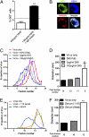
Inhibition of immune-complex internalization is not due to FcγR competition but due to increased immune-complex size. (A) Percentage of DiD+ cells at 30 min post uptake when in complex with 400 μg/mL h3H5 or 1.56 μg/mL h3H5 with addition of 398.44 μg/mL of human isotype control. (B) Subcellular localization of DENV-2 complexed with 1.56 μg/mL h3H5 and with the addition of 398.44 μg/mL human IgG1 isotype control. LAMP-1 is green, DiD-labeled DENV-2 is blue, and human antibodies are red. (Scale bar, 7.5 μm.) (C) Proportion of total viral RNA extracted from the various sucrose fractions. Proportion of viral RNA in each fraction was determined by dividing the viral RNA copy number in that fraction by the viral RNA copy number in the entire gradient. Shown are the moving averages of free virus and 33.3-μg/mL Fab fragments of h3H5 or virus in complex with 3 μg/mL h3H5 or with 100 μg/mL h3H5 using qPCR. (D) Diameter of the immune complexes measured using dynamic light scattering in the respective sucrose fractions with peak viral RNA copy number. (E and F) Similar to C and D, but with 1:10 or undiluted serum. Data are represented as mean ± SD. **P < 0.01.
Fig. 4.
Fig. 4.
FcγRIIB is involved in the inhibition of immune-complex internalization of larger viral aggregates. (A) THP-1 cells exposed to media (mock), DENV-2 in complex with 1.56 μg/mL h3H5, and DENV-2 complexed with 100 μg/mL h3H5 30 min post infection. Cell lysates were immunoblotted with anti-SHP-1 and anti-phospho-SHP-1 (p-SHP-1). (B) THP-1 cells transfected with a control siRNA or siRNA against FcγRIIB. Cell lysates were immunoblotted with anti-FcγRIIB, anti-FcγRI, anti-FcγRIIA, and LAMP-1 antibodies. LAMP-1 served as a loading control. (C) Percentage of internalized DiD-labeled DENV-2 in THP-1 cells transfected with control siRNA or siRNA against FcγRIIB when in complex with various h3H5 concentrations. Dashed line indicates DiD+ cells in the presence of DiD-labeled DENV-2 only, without antibodies. (D) THP-1 cells subjected to mock transfection or transfected with FcγRIIB DNA. Cell lysates were immunoblotted with anti-FcγRIIB, anti-FcγRI, anti-FcγRIIA, and LAMP-1 antibodies (loading control). (E) Same as C, but using THP-1 cells with mock transfection or with transfected FcγRIIB DNA. (F) Plaque titers at 72 h post infection on cells with mock transfection or with overexpression of FcγRIIB. Dashed line indicates plaque titers in the presence of DENV-2 only, without h3H5. No significant differences between control and treated cells were observed with virus-only infection. Data are represented as mean ± SD.

Similar articles

Cited by

References

    1. Kyle JL, Harris E. Global spread and persistence of dengue. Annu Rev Microbiol. 2008;62:71–92. - PubMed
    1. Wilder-Smith A, Ooi EE, Vasudevan SG, Gubler DJ. Update on dengue: Epidemiology, virus evolution, antiviral drugs, and vaccine development. Curr Infect Dis Rep. 2010;12:157–164. - PubMed
    1. Simmons CP, et al. Maternal antibody and viral factors in the pathogenesis of dengue virus in infants. J Infect Dis. 2007;196:416–424. - PMC - PubMed
    1. Halstead SB, O'Rourke EJ. Dengue viruses and mononuclear phagocytes. I. Infection enhancement by non-neutralizing antibody. J Exp Med. 1977;146:201–217. - PMC - PubMed
    1. Halstead SB. Pathogenesis of dengue: Challenges to molecular biology. Science. 1988;239:476–481. - PubMed

Publication types