. 2011 Jul 19;108(29):11842-7.
doi: 10.1073/pnas.1101791108. Epub 2011 Jul 11.

Humanized mice with ectopic artificial liver tissues

Affiliations

Humanized mice with ectopic artificial liver tissues

Alice A Chen et al. Proc Natl Acad Sci U S A. .

Abstract

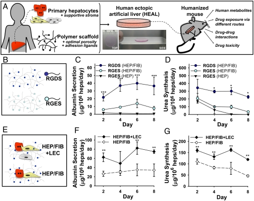
"Humanized" mice offer a window into aspects of human physiology that are otherwise inaccessible. The best available methods for liver humanization rely on cell transplantation into immunodeficient mice with liver injury but these methods have not gained widespread use due to the duration and variability of hepatocyte repopulation. In light of the significant progress that has been achieved in clinical cell transplantation through tissue engineering, we sought to develop a humanized mouse model based on the facile and ectopic implantation of a tissue-engineered human liver. These human ectopic artificial livers (HEALs) stabilize the function of cryopreserved primary human hepatocytes through juxtacrine and paracrine signals in polymeric scaffolds. In contrast to current methods, HEALs can be efficiently established in immunocompetent mice with normal liver function. Mice transplanted with HEALs exhibit humanized liver functions persistent for weeks, including synthesis of human proteins, human drug metabolism, drug-drug interaction, and drug-induced liver injury. Here, mice with HEALs are used to predict the disproportionate metabolism and toxicity of "major" human metabolites using multiple routes of administration and monitoring. These advances may enable manufacturing of reproducible in vivo models for diverse drug development and research applications.


Conflict of interest statement

The authors declare no conflict of interest.

Figures

Fig. 1.
Fig. 1.
Human ectopic artificial livers. (A) Schematic depicting the fabrication, implantation, and utility of HEALs for humanizing mice. Primary hepatocytes are cocultivated with stabilizing stromal fibroblasts on collagen-coated plates for 7–10 d, then encapsulated with liver endothelial cells in PEG-DA (20 kDa, 10% wt/vol) scaffolds derivatized with adhesion peptides. The resulting HEAL is approximately 20-mm diameter and 250-μm thick and comprises ∼0.5 × 106 human hepatocytes (inset shows a 10-mm-diameter version from top and side views). After implantation into laboratory mice, engrafted and vascularized HEALs establish humanized models for drug development applications. (BD) Covalent modification of PEG-DA hydrogels with peptide sequences RGDS (ligand) or RGES (negative control) (10 μmol/mL), and assessment of encapsulated primary HEP or HEP/FIB for albumin secretion (C) and urea synthesis (D) over time. (E and F) Coencapsulation of LEC with HEP/FIB cocultures in RGDS-derivatized PEG-DA hydrogels, and assessment of albumin secretion (E) and urea synthesis (F) over time. *p < 0.01, **p < 0.05, ***p < 0.001 for n = 6 and SEM.
Fig. 2.
Fig. 2.
Characterization of HEALs for drug metabolism gene expression and functions. (A) Heat map displays of LMA-Luminex analysis for 83 human-specific drug metabolism genes and transcription factors, shown separately for independent experiments analyzing different hepatocyte donors (donor A or B). Columns represent replicate loadings of RNA extracted from 2D HEP/FIB cultures (“2D”) or 3D HEP/FIB HEALs (“3D”) on day 10 postencapsulation. mRNA expression is determined relative to average of control gene transferrin, and heat maps are row-normalized to distinguish relative 2D to 3D differences. (B) Select gene sets comparing the relative mRNA expression of 3D HEP/FIB HEALs (open bars), normalized to 2D HEP/FIB cultures (filled bars) for phase I, nuclear receptors, phase II, and phase III drug metabolism genes after DMSO exposure. Data represent the mean and SEM of Luminex-loaded replicates for donor A (black) and donor B (gray). (C) CYP450 activity, induction, and drug interactions in day 6 3D HEP/FIB+LEC HEALs from donor A, treated with clinical inducers OME (40 μM) or RIF (25 μM) in vitro. Cultures were treated daily with inducers for 3 d before incubation with ER or TEST, conventional substrates for CYP1A2 and 3A4, respectively. Fold-induction of CYP450 activity was determined by normalization to DMSO control. By Student’s t test, *p < 0.01, ***p < 0.001 for n = 4 and SD.
Fig. 3.
Fig. 3.
Humanized mice via ectopic implantation of HEALs and functional assessment in vivo. (A) In vivo bioluminescence imaging and (B) quantification of reporter albumin-firefly luciferase (Fluc) HEALs implanted in the intraperitoneal cavity (IP) or subcutaneous space (SC) of athymic nude mice (n = 8 for IP, n = 3 for SC implants). Reporter albumin-Fluc HEALs were fabricated using HEP/FIB cocultures transduced by lentivirus to express luciferase under the human albumin promoter, prior to encapsulation with LEC (HEP/FIB + LEC). (C) Human serum albumin detected in mice humanized with intraperitoneal HEALs. Red bars mark average human serum albumin levels at each timepoint for n = 6 to 8 mice. (D) Representative photographs of mice with HEAL, perfused with yellow Microfil silicone rubber on day 35 after implantation. HEAL is shown pseudooutlined and exposed within peritoneal cavity (arrow; Left), and partially dissected (Right). (E) Micro-CT angiography scan and maximum intensity projection of representative extracted HEAL. (F) Quantification of vascular volume in extracted HEAL, based on 30-μm micro-CT slices from surface interfacing with host mesentery. The dashed line marks the expected opposite boundary surface of the HEAL based on its fabricated thickness of approximately 250 μm. (Scale bars: 5, 2, 5 mm.)
Fig. 4.
Fig. 4.
Humanized mice using different donors or recipients. Drug dosing via multiple routes of administration and prediction of major human metabolites, drug–drug interactions, and toxicity. (A) Representative micrographs of cryopreserved primary hepatocytes from donors A and B, cocultivated with stromal fibroblasts for 7 d prior to 3D encapsulation. (Scale bar: 75 μm.) (B) Characterization of donors A- or B-derived HEALs for CYP2D6 activity by exposure to CYP2D6 substrate debrisoquine and quantification of debrisoquine hydroxylation. (C) In vivo bioluminescence imaging and quantification of reporter albumin-firefly luciferase HEALs from donor A, implanted in the intraperitoneal cavity of athymic nude, immunocompetent C57/BL6, or Swiss–Webster mice. Representative images 3 d after implantation are shown. (D) Pharmacokinetic analysis of serum metabolite 7-HC formation in humanized nude mice exposed to coumarin via i.p. injection (Upper), and serum metabolite 4-OHDB formation in humanized C57/Bl6 mice exposed to debrisoquine via oral gavage (Lower). (E) Detection of urinary 7-HC (Upper) or 4-OHDB (Lower) metabolite excretion over 4 h in vivo. (F) Identification of major human metabolites based on the metabolic ratio (MR; parent AUC over metabolite AUC). Red dashed line represents the lower threshold for classification as a major metabolite (0.1 MR). (G) Timeline of drug–drug interaction (DDI) study. Humanized mice (n = 6 per group) were administered RIF (25 mg/kg) daily for 3 d before (H) extraction of HEAL and incubation with CYP1A2 substrate ER or CYP3A4 substrate TEST ex vivo, or (I) in vivo oral exposure to APAP and serum assessment of human albumin production. Fold-induction of CYP450 activity was determined by normalization to DMSO. *p < 0.05, **p < 0.01, ***p < 0.001 for n as indicated and SEM. Error bars are SEM for n = 3 or greater.

Comment in

  • Mice with artificial human liver.
    Kamiya A, Miyajima A. Kamiya A, et al. Hepatology. 2012 Mar;55(3):974-6. doi: 10.1002/hep.25503. Hepatology. 2012. PMID: 22362602 No abstract available.

Similar articles

Cited by

References

    1. Huebsch N, Mooney DJ. Inspiration and application in the evolution of biomaterials. Nature. 2009;462:426–432. - PMC - PubMed
    1. Ploss A, et al. Persistent hepatitis C virus infection in microscale primary human hepatocyte cultures. Proc Natl Acad Sci USA. 2010;107:3141–3145. - PMC - PubMed
    1. Khetani SR, Bhatia SN. Microscale culture of human liver cells for drug development. Nat Biotechnol. 2008;26:120–126. - PubMed
    1. Sung JH, Kam C, Shuler ML. A microfluidic device for a pharmacokinetic-pharmacodynamic (PK-PD) model on a chip. Lab Chip. 2010;10:446–455. - PubMed
    1. Shultz LD, Ishikawa F, Greiner DL. Humanized mice in translational biomedical research. Nat Rev Immunol. 2007;7:118–130. - PubMed

Publication types