. 2011 Jul 26;108(30):12407-12.
doi: 10.1073/pnas.1104295108. Epub 2011 Jul 11.

Targeted Sos1 deletion reveals its critical role in early T-cell development

Affiliations

Targeted Sos1 deletion reveals its critical role in early T-cell development

Robert L Kortum et al. Proc Natl Acad Sci U S A. .

Abstract

Activation of the small G protein Ras is required for thymocyte differentiation. In thymocytes, Ras is activated by the Ras guanine exchange factors (RasGEFs) Sos1, Sos2, and RasGRP1. We report the development of a floxed allele of sos1 to assess the role of Sos1 during thymocyte development. Sos1 was required for pre-T-cell receptor (pre-TCR)- but not TCR-stimulated developmental signals. Sos1 deletion led to a partial block at the DN-to-DP transition. Sos1-deficient thymocytes showed reduced pre-TCR-stimulated proliferation, differentiation, and ERK phosphorylation. In contrast, TCR-stimulated positive selection, and negative selection under strong stimulatory conditions, remained intact in Sos1-deficient mice. Comparison of RasGEF expression at different developmental stages showed that relative to Sos2 and RasGRP1, Sos1 is most abundant in DN thymocytes, but least abundant in DP thymocytes. These data reveal that Sos1 is uniquely positioned to affect signal transduction early in thymocyte development.


Conflict of interest statement

The authors declare no conflict of interest.

Figures
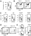
Fig. 1.
Fig. 1.
T-cell–specific deletion of Sos1 impairs early thymocyte development. (A) Total numbers of thymocytes isolated from 8-wk-old Sos1+/+ (n = 10) and Sos1−/−(T) (n = 18) mice. Each diamond denotes an individual mouse, and the bar denotes the average for the group. ***P < 0.001. (B) Flow cytometry dot plots of total thymocytes stained with anti-CD4 and anti-CD8 from representative 8-wk-old Sos1+/+ and Sos1−/−(T) mice from A. (C) Total numbers of DN, DP, CD4SP, and CD8SP thymocytes from A and B. **P < 0.01, ***P < 0.001. (D) Flow cytometry dot plots of gated lin (CD4CD8CD11bTCRβTCRγδTer119B220NK1.1; SI Materials and Methods) DN thymocytes stained with anti-CD44 and anti-CD25 to identify DN1–DN4 thymocytes from 8-wk-old Sos1+/+ (n = 9) and Sos1−/−(T) (n = 10) mice. (E) Total numbers of linCD44CD25+ (DN3) thymocytes, linCD44CD25 (DN4) thymocytes, and the DN3:DN4 ratio from D. **P < 0.01.
Fig. 2.
Fig. 2.
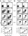
Sos1 is required for anti-CD3ε–induced proliferation and differentiation in Rag2−/− thymocytes. (A and B) Flow cytometry dot plots of total thymocytes stained with anti-CD4 and anti-CD8 (Upper) and quantification of total thymocyte counts (Lower) 0, 3, 5, or 7 d following a single i.p. injection of 100 μg anti-CD3ε antibody (2C11) from (A) Sos1+/+Rag2−/− (black squares) and Sos1−/−(T)Rag2−/− (gray diamonds) or (B) RasGRP1+/+Rag2−/− (black squares) and RasGRP1−/−Rag2−/− (gray diamonds). n = 3 for each time point. Data represent mean ± SD.
Fig. 3.
Fig. 3.
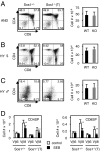
Sos1 is not required during DP thymocyte selection. (A–C) Flow cytometry dot plots of total thymocytes stained with anti-CD4 and anti-CD8 (Left) and quantification of total thymocyte numbers (Right) from 8-wk-old TCR transgenic Sos1+/+ and Sos1−/−(T) mice. n ≥ 4 for each group. Data represent mean ± SD. (A) AND+ mice, (B) HY+ female mice, (C) HY+ male mice. (D) Total numbers of Vβ6+ or Vβ8+ CD4SP and CD8SP thymocytes from Sos1+/+ and Sos1−/−(T) mice injected with PBS (white) or SEB (black) as described in SI Materials and Methods. n = 7 for each group. Data represent mean ± SD. **P < 0.01.
Fig. 4.
Fig. 4.
Sos1 is preferentially required for signal transduction early in thymocyte development. (A–C) Western blotting for phospho-ERK and total ERK (Left, quantified Right) in (A) total thymocytes from Sos1+/+Rag2−/− vs. Sos1−/−(T)Rag2−/− mice stimulated with 25 μg/mL anti-CD3ε antibody, (B) purified DP thymocytes from Sos1+/+ vs. Sos1−/−(T) mice stimulated with 10 μg/mL anti-CD3ε antibody, and (C) purified CD4+ LN cells from Sos1f/f vs. Sos1f/f;CD4-cre+ mice stimulated with 10 μg/mL anti-CD3ε antibody. All stimulations were for the indicated times in minutes. Data represent mean ± SD from two independent experiments.
Fig. 5.
Fig. 5.
Sos1 is preferentially expressed early in thymocyte development. Western blotting (Left) and quantification (Right) for Sos1, Sos2, RasGRP1, and β-actin in DN3, DP, and CD4+ LN T cells purified from Rag2−/− thymus, B6 thymus, or B6 LN, respectively. Each lane represents one of three independently isolated populations. Data represent mean ± SD.

Similar articles

Cited by

References

    1. Buday L, Downward J. Many faces of Ras activation. Biochim Biophys Acta. 2008;1786:178–187. - PubMed
    1. Hanahan D, Weinberg RA. The hallmarks of cancer. Cell. 2000;100:57–70. - PubMed
    1. Mor A, Philips MR. Compartmentalized Ras/MAPK signaling. Annu Rev Immunol. 2006;24:771–800. - PubMed
    1. Daniels MA, et al. Thymic selection threshold defined by compartmentalization of Ras/MAPK signalling. Nature. 2006;444:724–729. - PubMed
    1. Swat W, Shinkai Y, Cheng HL, Davidson L, Alt FW. Activated Ras signals differentiation and expansion of CD4+8+ thymocytes. Proc Natl Acad Sci USA. 1996;93:4683–4687. - PMC - PubMed

Publication types

LinkOut - more resources