. 2011 Oct;55(10):4774-81.
doi: 10.1128/AAC.00151-11. Epub 2011 Jul 11.

Diphenylpyrazole-derived compounds increase survival time of mice after prion infection

Affiliations

Diphenylpyrazole-derived compounds increase survival time of mice after prion infection

Fabienne Leidel et al. Antimicrob Agents Chemother. 2011 Oct.

Abstract

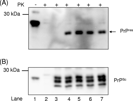
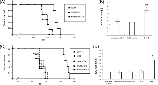
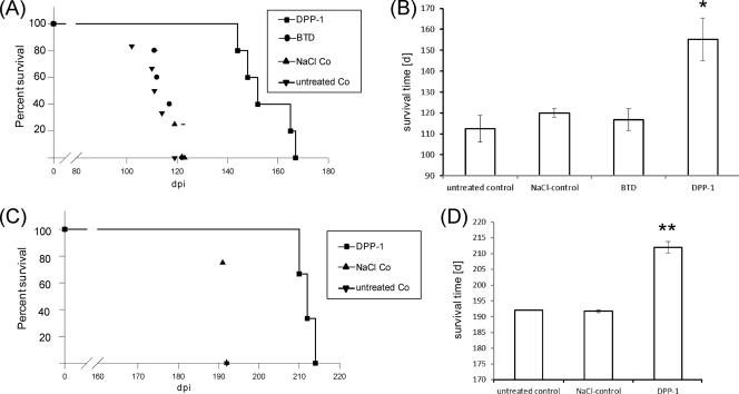
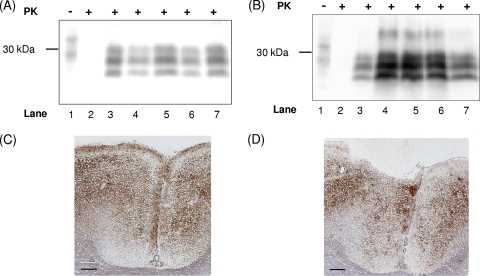
Transmissible spongiform encephalopathies (TSEs) represent a group of fatal neurodegenerative disorders that can be transmitted by natural infection or inoculation. TSEs include scrapie in sheep, bovine spongiform encephalopathy (BSE) in cattle, and Creutzfeldt-Jakob disease (CJD) in humans. The emergence of a variant form of CJD (vCJD), which has been associated with BSE, produced strong pressure to search for effective treatments with new drugs. Up to now, however, TSEs have proved incurable, although many efforts have been made both in vitro and in vivo to search for potent therapeutic and prophylactic compounds. For this purpose, we analyzed a compound library consisting of 10,000 compounds with a cell-based high-throughput screening assay dealing with scrapie-infected scrapie mouse brain and ScN(2)A cells and identified a new class of inhibitors consisting of 3,5-diphenylpyrazole (DPP) derivatives. The most effective DPP derivative showed half-maximal inhibition of PrP(Sc) formation at concentrations (IC(50)) of 0.6 and 1.2 μM, respectively. This compound was subsequently subjected to a number of animal experiments using scrapie-infected wild-type C57BL/6 and transgenic Tga20 mice. The DPP derivative induced a significant increase of incubation time both in therapeutic and prophylactic experiments. The onset of the prion disease was delayed by 37 days after intraperitoneal and 42 days after oral application, respectively. In summary, we demonstrate a high in vitro efficiency of DPP derivatives against prion infections that was substantiated in vivo for one of these compounds. These results indicate that the novel class of DPP compounds should comprise excellent candidates for future therapeutic studies.


Figures

Fig. 1.
Fig. 1.
DPP derivatives do not interfere with cell-free PrPres formation and do not dissolve existing PrPSc aggregates. (A) Controls were performed as follows: PrPC in a 1:10 dilution without PrPSc or PK (lane 1), PrPC without PrPSc incubated with PK (lane 2), and PrPC incubated with PrPSc but stopped immediately by freezing (t0 control, lane 3). Lane 4 shows the PrPres fragments after incubation for 72 h with scrapie strain Me7 in the presence of 100 μM DPP-1 (lane 5), 100 μM DPP-2 (lane 6), or 100 μM DPP-3 (lane 7). Detection of PrPC and PrPres fragments was carried out with MAb 3F4. (B) The membrane in panel A was stripped and reincubated with PAb Ra10 for the detection of PrPSc seeds in lanes 3 to 7.
Fig. 2.
Fig. 2.
DPP-1 prolongs the incubation time in scrapie-infected mice after i.p. treatment. (A) Primary efficiency test. Kaplan-Meier survival analysis of i.c. scrapie-infected C57BL/6 mice was performed after i.p. treatment with DPP-1. The treatment groups included untreated controls (▾), DMSO controls (▴), and DPP-1-treated mice (■). (B) Mean survival times ± the standard deviations as determined by the primary efficiency test. Comparison of DPP-1 versus untreated controls and DMSO controls was carried out by using the log-rank test (**, P < 0.01). (C) Intraperitoneal therapy. Kaplan-Meier survival analysis of i.p. scrapie-infected Tga20 mice was performed after i.p. treatment with BTD or DPP-1. The treatment groups included untreated controls (▾), vehicle controls (▴), DMSO controls (◆), benzothiazol (BTD)-treated mice (●), and DPP-1-treated mice (■). (D) Mean survival times in i.p. therapy ± the standard deviation. Comparison of DPP-1-treated versus untreated controls, DMSO controls, vehicle control, and BTD-treated mice was carried out by using the log-rank test (*, P < 0.05).
Fig. 3.
Fig. 3.
DPP-1 prolongs incubation time in scrapie-infected mice after oral treatment. (A) Oral treatment. Kaplan-Meier survival analysis of i.p. scrapie-infected Tga20 mice was performed after oral treatment with DPP-1 or benzothiazol (BTD). The treatment groups included untreated controls (▾), NaCl controls (▴), benzothiazol (BTD)-treated mice (●), and DPP-1-treated mice (■). (B) Mean survival times in primary efficiency test ± the standard deviation. Comparison of DPP-1 versus untreated controls, NaCl controls, and BTD-treated mice was carried out by using the log-rank test (*, P < 0.05). (C) Oral prophylaxis. Kaplan-Meier survival analysis of i.p. scrapie-infected C57BL/6 mice was performed after oral treatment with DPP-1. The treatment groups included untreated controls (▾), NaCl controls (▴) and DPP-1-treated mice (■). (D) Mean survival times in oral prophylaxis ± the standard deviation. Comparison of DPP-1-treated versus untreated controls and NaCl controls was carried out by using the log-rank test (**, P < 0.01).
Fig. 4.
Fig. 4.
PrPSc accumulation in the brains of selected mice challenged with RML. Immunoblot analysis of RML-infected mouse brain homogenate. (A) Control experiments were carried out using PrPC without PK (lane 1), PrPC digested with PK (lane 2), or PrPSc digested with PK (lane 3). Lanes 4 and 5 show PrPSc fragments of mice treated with osmotic pumps infused with DPP-1 (lane 4) or DMSO control (lane 5). Lanes 6 and 7 display PK-digested PrPSc fragments from orally treated mice (postinoculation) either with (lane 6) or without (lane 7) DPP-1. (B) Controls were established similar to those in Fig. 4A: PrPC without K (lane 1), PrPC digested with PK (lane 2), or PrPSc digested with PK (lane 3). Lanes 4 and 5 show PrPSc fragments derived from i.c.-inoculated mice treated with DPP-1 (lane 4) or not treated (lane 5). Lanes 6 and 7 display PK-digested PrPSc fragments of i.p.-infected mice after oral prophylactic administration of DPP-1 (lane 6) compared to the NaCl controls (lane 7). Detection of PrPC or PrPSc was carried out with MAb SAF70 and MAb RGS-His for the detection of molecular mass markers. PrPSc immunohistochemistry of brain tissue was performed. (C and D) Cortices of DPP-1-treated (C) and untreated (D) i.p.-infected C57BL/6 mice (oral prophylaxis) at the terminal stage of disease. MAb ICSM 18, 1:250; bar, 200 μm.

Similar articles

Cited by

References

    1. Adjou K. T., Seman M. 2002. What are the prospects for pharmacological treatment of prion disease? Therapie 57:123–127 - PubMed
    1. Andrews N. J., et al. 2003. Deaths from variant Creutzfeldt-Jakob disease in the UK. Lancet 361:751–752 - PubMed
    1. Ansar S., et al. 2007. A non-toxic Hsp90 inhibitor protects neurons from Aβ-induced toxicity. Bioorg. Med. Chem. Lett. 17:1984–1990 - PubMed
    1. Birkett C. R., et al. 2001. Scrapie strains maintain biological phenotypes on propagation in a cell line in culture. EMBO J. 20:3351–3358 - PMC - PubMed
    1. Brown D. R. 2002. Mayhem of the multiple mechanisms: modeling neurodegeneration in prion disease. J. Neurochem. 58:1720–1725 - PubMed

Publication types