. 2011 Aug 15;187(4):1807-15.
doi: 10.4049/jimmunol.1100296. Epub 2011 Jul 11.

The Src family kinase Fgr is critical for activation of mast cells and IgE-mediated anaphylaxis in mice

Affiliations

The Src family kinase Fgr is critical for activation of mast cells and IgE-mediated anaphylaxis in mice

Jun Ho Lee et al. J Immunol. .

Abstract

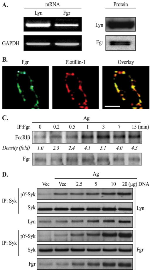
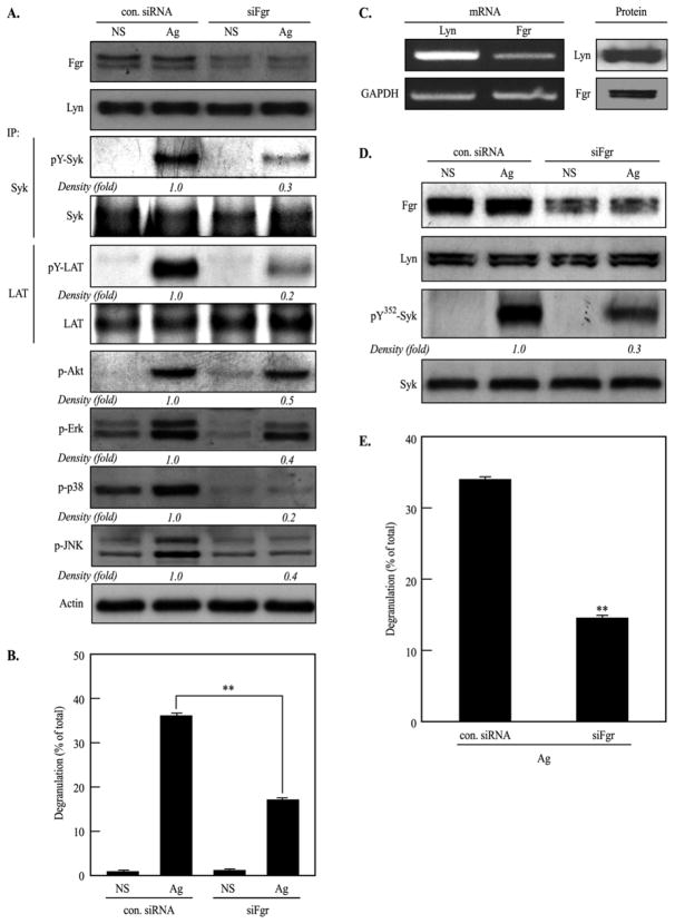
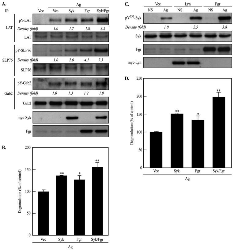
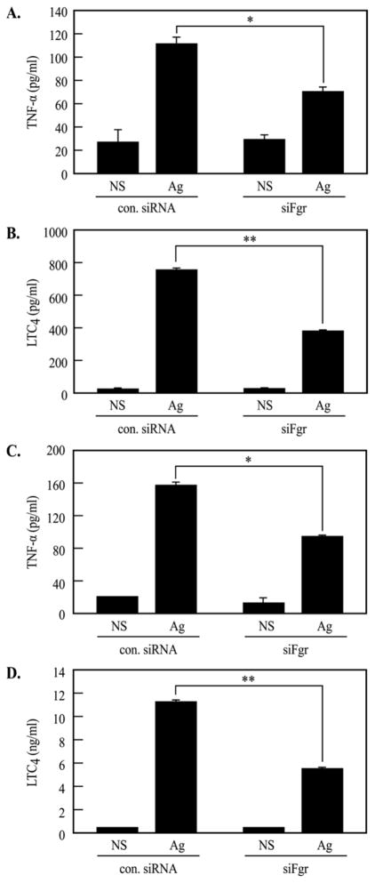
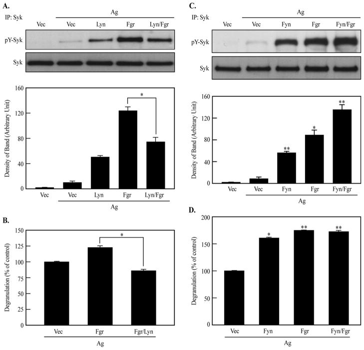
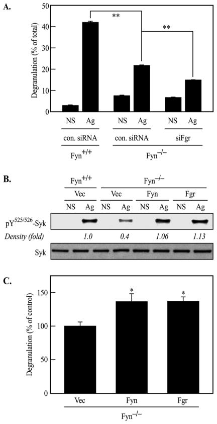
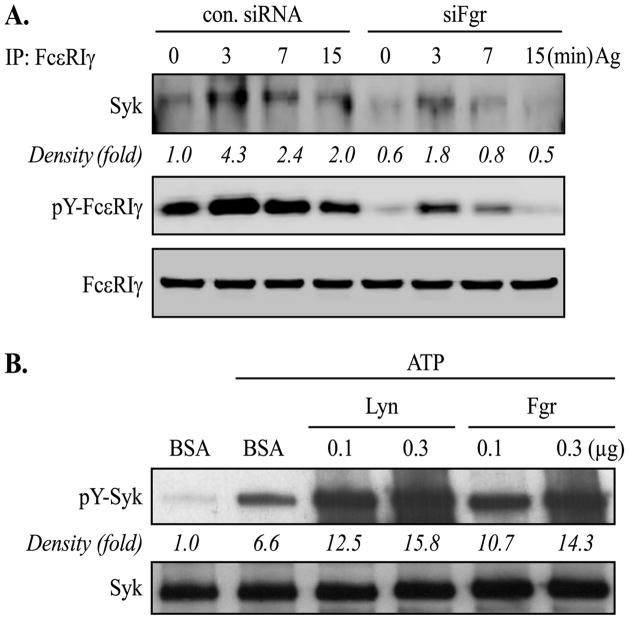
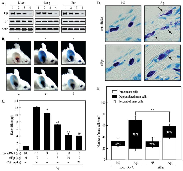
Mast cells are critical for various allergic disorders. Mast cells express Src family kinases, which relay positive and negative regulatory signals by Ag. Lyn, for example, initiates activating signaling events, but it also induces inhibitory signals. Fyn and Hck are reported to be positive regulators, but little is known about the roles of other Src kinases, including Fgr, in mast cells. In this study, we define the role of Fgr. Endogenous Fgr associates with FcεRI and promotes phosphorylation of Syk, Syk substrates, which include linkers for activation of T cells, SLP76, and Gab2, and downstream targets such as Akt and the MAPKs in Ag-stimulated mast cells. As a consequence, Fgr positively regulates degranulation, production of eicosanoids, and cytokines. Fgr and Fyn appeared to act in concert, as phosphorylation of Syk and degranulation are enhanced by overexpression of Fgr and further augmented by overexpression of Fyn but are suppressed by overexpression of Lyn. Moreover, knockdown of Fgr by small interfering RNAs (siRNAs) further suppressed degranulation in Fyn-deficient bone marrow-derived mast cells. Overexpression of Fyn or Fgr restored phosphorylation of Syk and partially restored degranulation in Fyn-deficient cells. Additionally, knockdown of Fgr by siRNAs inhibited association of Syk with FcεRIγ as well as the tyrosine phosphorylation of FcεRIγ. Of note, the injection of Fgr siRNAs diminished the protein level of Fgr in mice and simultaneously inhibited IgE-mediated anaphylaxis. In conclusion, Fgr positively regulates mast cell through activation of Syk. These findings help clarify the interplay among Src family kinases and identify Fgr as a potential therapeutic target for allergic diseases.


Conflict of interest statement

Disclosures

The authors have no financial conflict of interest.

Figures

FIGURE 1
FIGURE 1
Fgr is associated with the plasma membrane, interacts with FcεRI, and phosphorylates Syk in RBL-2H3 cells. A, left panel, Expression of message for Lyn, Fgr, and GAPDH was determined by RT-PCR. A, right panel, Whole cell lysates were subjected to immunoblot analysis. B, Fgr and Flotillin-1 were visualized in RBL-2H3 cells by confocal microscopy: scale bar, 10 μm. C, Fgr was immunoprecipitated (IP) and subjected to immunoblot analysis in RBL-2H3 cells stimulated with antigen (Ag). D, RBL-2H3 cells were transiently transfected with DNA constructs encoding vector (Vec), Lyn or Fgr as indicated. The cells were stimulated by antigen (Ag) or not. The level of overexpressed Lyn, Fgr or immunoprecipitated (IP) Syk from whole cell lysates were subjected to immunoblot analysis. Representative results from three or more independent experiments are shown.
FIGURE 2
FIGURE 2
Knockdown of Fgr suppresses Syk, Syk-dependent signals, and degranulation. RBL-2H3 cells (A and B) or BMMCs (D and E) were transiently transfected with siRNAs directed against Fgr (siFgr), or with the control siRNAs (con. siRNA), respectively. The cells were stimulated with antigen (Ag) or not stimulated (NS) for immunoblot analysis and measurement of degranulation. Immunoprecipitates (IP) for Syk and LAT or whole cell lysates were subjected to immunoblot analysis (A and D). C, left panel, Expression of message for Lyn, Fgr, and GAPDH was determined by RT-PCR in BMMCs. C, right panel, Whole cell lysates from BMMCs were subjected to immunoblot analysis. Representative images from three independent experiments are shown. (B and E) Cells were stimulated with antigen (Ag) for 15 min or not (NS) to measure release of the granule marker, β-hexosaminidase. Values for degranulation are the means ± SEM of values from three independent experiments: **P < 0.01.
FIGURE 3
FIGURE 3
Overexpression Fgr stimulates Syk-dependent signals and degranulation. RBL-2H3 cells (A–B) or BMMCs (C–D) were transiently transfected with DNA constructs encoding myc-Syk, Lyn, Fgr, or vector (Vec). A and C, Cells were stimulated with antigen (Ag) for 7 min or not stimulated (NS). Immunoprecipitates (IP) for LAT, SLP76, and Gab2 or whole cell lysates were subjected to immunoblot analysis. Representative results from three independent experiments are shown. B and D, Degranulation was determined. Values are the means ± SEM of values from three independent experiments: *P < 0.05 and **P < 0.01.
FIGURE 4
FIGURE 4
Transfection of the Fgr siRNAs impairs production of TNF-α and LTC4. RBL-2H3 cells (A and B) or BMMCs (C and D) were transfected with Fgr siRNAs (siFgr), or the control siRNAs (con. siRNA). IgE-primed cells were stimulated with antigen (Ag) for 8 h or not stimulated (NS) for analysis of TNF-α or LTC4 in culture media by ELISA. Values are the means ± the SEM of values from three independent experiments: *P < 0.05 and **P < 0.01.
FIGURE 5
FIGURE 5
Positive role of Fgr on phosphorylation of Syk is negated by overexpression of Lyn, but reinforced by Fyn. RBL-2H3 cells were transfected with individual or a combination of plasmids at suboptimal dose (3 μg each) encoding Lyn, Fgr, Fyn or Vector (Vec). A and C, IgE-primed cells were stimulated with antigen (Ag) for 7 min or not stimulated. Immunoprecipitates (IP) for Syk were analyzed by immunoblot analysis. Representative images (upper panels) and density of bands for phoshorylated Syk (lower panels) are the mean ± SEM from three independent experiments. B and D, IgE-primed cells were stimulated with antigen (Ag) for 15 min to measure degranulation. Values are the means ± the SEM of values from three independent experiments: *P < 0.05 and **P < 0.01.
FIGURE 6
FIGURE 6
Overexpression of Fgr or Fyn restores phosphorylation of Syk and partly so degranulation in Fyn-deficient cells. A, BMMCs isolated from Fyn+/+ or Fyn−/− C57BL6 mice were transfected with Fgr siRNAs (siFgr) or the control siRNAs (con. siRNA). IgE-primed cells were stimulated with antigen (Ag) for 15 min or not stimulated (NS) to measure degranulation. B, Fyn+/+ or Fyn−/− BMMCs were transfected with vector (Vec), Fgr, or Fyn DNA as indicated. Cells were primed with IgE and then stimulated with antigen (Ag) for 7 min or not (NS). Whole cell lysates were subjected to immunoblot analysis for detection of phosphorylated forms of Syk. Representative results from three independent experiments are shown. C, Fyn−/− BMMCs were transfected with vector (Vec), Fgr or Fyn DNA. IgE-primed cells were stimulated with antigen for 15 min to measure degranulation. Values are the means ± the SEM of values from three independent experiments: *P < 0.05 and **P < 0.01.
FIGURE 7
FIGURE 7
Fgr phosphorylates Syk in vitro and is also critical for recruitment of Syk to the γ subunit of FcεRI in antigen-stimulated cells. A, BMMCs were primed with IgE and then stimulated with antigen (Ag) for indicated times. The γ subunit of FcεRI (FcεRIγ) was immunoprecipitated (IP) from cell lysates with anti-FcεRIγ antibody, and precipitated proteins were subjected to immunoblot analysis with antibodies against Syk, FcεRIγ, and phosphotyrosine residues (pY) (4G10). B, Recombinant Syk was incubated with 100–300 ng of Lyn, Fgr, or 300 ng of bovine serum albumin (BSA) as a control protein in a kinase reaction buffer. Phosphorylated Syk was measured by immunoblot analysis. Representative images were obtained from three independent experiments.
FIGURE 8
FIGURE 8
Fgr is critical for IgE-mediated PCA reaction and degranulation in mice. The Fgr siRNAs (siFgr) that were used in vitro (Fig. 2D) were injected into BALB/c mice three times every 24 h before assessing the levels of Fgr protein in tissues and the PCA reaction as described in “Materials and Methods”. A, Extracts from liver, lung, and ear were subjected to Western blotting analysis: 1, control (con.) siRNAs (10 μg); 2, siFgr (1 μg) + con. siRNAs (9 μg); 3, siFgr (3 μg) + con. siRNAs (7 μg); 4, siFgr (10 μg). Results are typical from at least three independent experiments. B, Representative photographs of ears (a, con. siRNAs (10 μg) without Ag; b, con. siRNAs (10 μg) with Ag; c, siFgr (1 μg) + con. siRNAs (9 μg) with Ag; d, siFgr (3 μg) + con. siRNAs (7 μg) with Ag; e, siFgr (10 μg) with Ag; f, Cetrizine (Cet, 20 mg/kg) with Ag). C, Quantitative data for ear-tissue content of Evans blue are expressed as the mean ± SEM of values from three independent experiments, each with 10 mice. The asterisks indicate significant difference from antigen (Ag)-stimulated controls with con. siRNAs (*p < 0.05; **p < 0.01). Cetirizine was orally administered 1 h before the antigen injection as a typical anti-histamine reference drug. D, Control siRNA or siFgr were administered (10 μg of each) as for panel B and the ear skins were prepared for histological examination as described in “Materials and Methods”. Representative histological images are shown: arrows indicate degranulated mast cells. E, The histograms show the percentage of degranulated mast cells in the given number of mast cells in ear skin sections. Values for degranulated mast cells are expressed as percent of total number of mast cells and are the means ± the SEM of values from three independent experiments: **P < 0.01.

Similar articles

Cited by

References

    1. Beaven MA. Our perception of the mast cell from Paul Ehrlich to now. Eur J Immunol. 2009;39:11–25. - PMC - PubMed
    1. Kalesnikoff J, Galli SJ. New developments in mast cell biology. Nat Immunol. 2008;9:1215–1223. - PMC - PubMed
    1. Bischoff SC. Role of mast cells in allergic and non-allergic immune responses: comparison of human and murine data. Nat Rev Immunol. 2007;7:93–104. - PubMed
    1. Gilfillan AM, Rivera J. The tyrosine kinase network regulating mast cell activation. Immunol Rev. 2009;228:149–169. - PMC - PubMed
    1. Odom S, Gomez G, Kovarova M, Furumoto Y, Ryan JJ, Wright HV, Gonzalez-Espinosa C, Hibbs ML, Harder KW, Rivera J. Negative Regulation of Immunoglobulin E-dependent Allergic Responses by Lyn Kinase. J Exp Med. 2004;199:1491–1502. - PMC - PubMed

Publication types

MeSH terms