

Association between in vivo fluorine 18-labeled flutemetamol amyloid positron emission tomography imaging and in vivo cerebral cortical histopathology
- PMID: 21747004
- PMCID: PMC4532383
- DOI: 10.1001/archneurol.2011.153
Association between in vivo fluorine 18-labeled flutemetamol amyloid positron emission tomography imaging and in vivo cerebral cortical histopathology
Abstract
Objective: To determine the correspondence of in vivo quantitative estimates of brain uptake of fluorine 18-labeled flutemetamol with immunohistochemical estimates of amyloid levels in patients who underwent previous biopsy.
Design: Cross-sectional study of ¹⁸F-flutemetamol positron emission tomography (PET) findings in patients with prior cortical biopsy specimen stained for the presence or absence of amyloid plaques.
Setting: University hospital. Patients Seven patients who previously had a prior right frontal cortical biopsy at the site of ventriculoperitoneal placement for presumed normal pressure hydrocephalus were recruited. Inclusion criteria included an adequate biopsy specimen for detection and quantification of β-amyloid pathology and age older than 50 years. Intervention All patients underwent an ¹⁸F-flutemetamol PET scan.
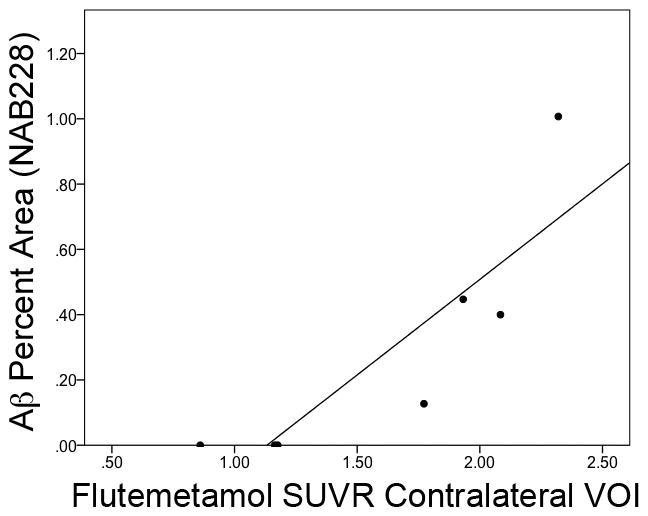
Main outcome measures: Quantitative measures of ¹⁸F-flutemetamol uptake (standardized uptake value ratio, a ratio of mean target cortex activity divided by that in a cerebellar reference region) were made at a location contralateral to the biopsy site and compared with estimates of amyloid load based on immunohistochemical and histological staining.
Results: There was complete agreement between visual reads of ¹⁸F-flutemetamol PET scans (3 blinded readers with majority rule) and histology. A regression model, including time from biopsy as a covariate, demonstrated a significant relationship (P = .01) between ¹⁸F-flutemetamol uptake and percentage of area of amyloid measured by a monoclonal antibody raised against amyloid (NAB228). Similar results were found with the amyloid-specific monoclonal antibody 4G8 and Thioflavin S.
Conclusion: To our knowledge, these data are the first to demonstrate the concordance of ¹⁸F-flutemetamol PET imaging with histopathology, supporting its sensitivity to detect amyloid and potential use in the study and detection of Alzheimer disease.
Figures


Comment in
-
Amyloid imaging: liberal or conservative? Let the data decide.Arch Neurol. 2011 Nov;68(11):1377-8. doi: 10.1001/archneurol.2011.152. Epub 2011 Jul 11. Arch Neurol. 2011. PMID: 21747005 No abstract available.
References
-
- Small GW, et al. PET of brain amyloid and tau in mild cognitive impairment. N Engl J Med. 2006;355(25):2652–2663. - PubMed
-
- Klunk WE, et al. Imaging brain amyloid in Alzheimer’s disease with Pittsburgh Compound-B. Ann Neurol. 2004;55(3):306–319. - PubMed
-
- Verhoeff NP, et al. In-vivo imaging of Alzheimer disease beta-amyloid with [11C]SB-13 PET. Am J Geriatr Psychiatry. 2004;12(6):584–595. - PubMed
-
- Rowe CC, et al. Imaging of amyloid beta in Alzheimer’s disease with 18F-BAY94-9172, a novel PET tracer: proof of mechanism. Lancet Neurol. 2008;7(2):129–135. - PubMed
-
- Lin KJ, et al. Whole-body biodistribution and brain PET imaging with [18F]AV-45, a novel amyloid imaging agent--a pilot study. Nucl Med Biol. 2010;37(4):497–508. - PubMed
Publication types
MeSH terms
Substances
Grants and funding
LinkOut - more resources
Full Text Sources
Medical
