. 2011 Aug 2;124(5):572-81.
doi: 10.1161/CIRCULATIONAHA.111.021352. Epub 2011 Jul 11.

Controlled and cardiac-restricted overexpression of the arginine vasopressin V1A receptor causes reversible left ventricular dysfunction through Gαq-mediated cell signaling

Affiliations

Controlled and cardiac-restricted overexpression of the arginine vasopressin V1A receptor causes reversible left ventricular dysfunction through Gαq-mediated cell signaling

Xue Li et al. Circulation. .

Abstract

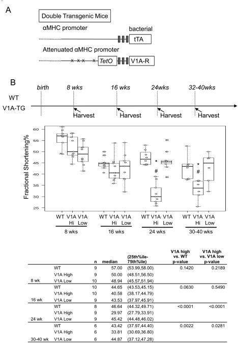
Background: [Arg8]-vasopressin (AVP) activates 3 G-protein-coupled receptors: V1A, V2, and V1B. The AVP-V1A receptor is the primary AVP receptor in the heart; however, its role in cardiac homeostasis is controversial. To better understand AVP-mediated signaling in the heart, we created a transgenic mouse with controlled overexpression of the V1A receptor.

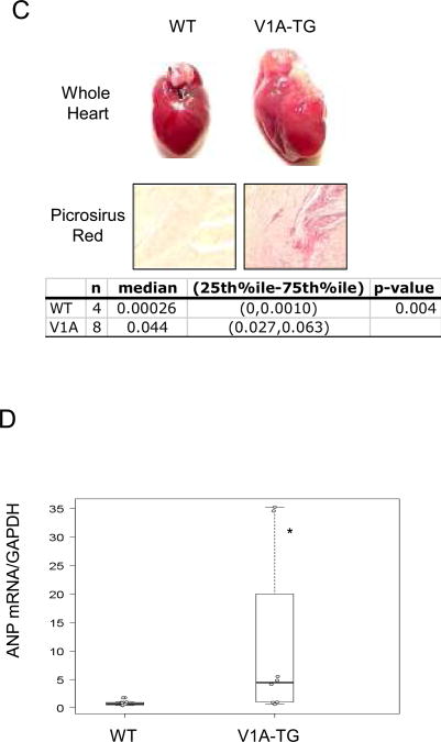
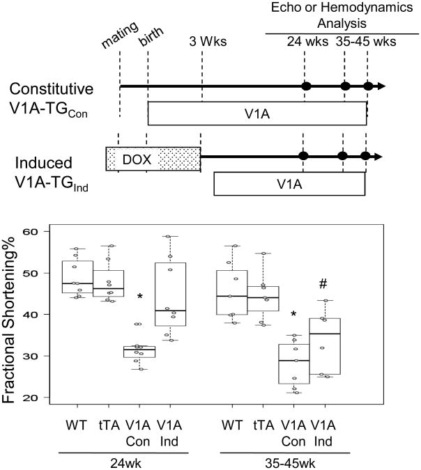
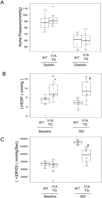
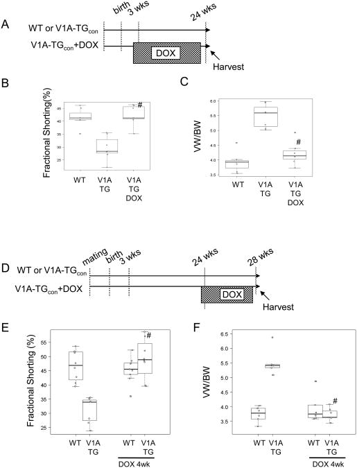
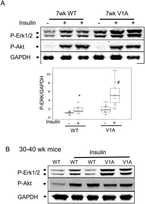
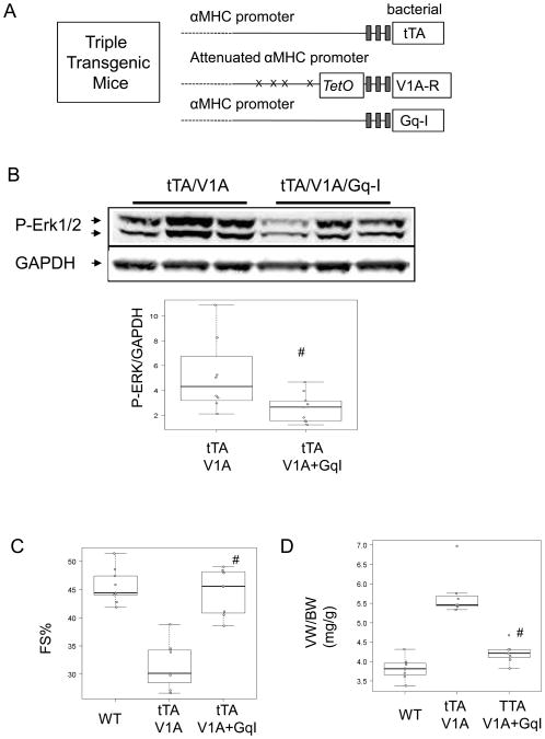
Methods and results: The V1A receptor transgene was placed under the control of the tetracycline-regulated, cardiac-specific α-myosin heavy chain promoter (V1A-TG). V1A-TG mice had a normal cardiac function phenotype at 10 weeks of age; however, by 24 weeks of age, tetracycline-transactivating factor/V1A-TG mouse hearts had reduced cardiac function, cardiac hypertrophy, and dilatation of the ventricular cavity. Contractile dysfunction was also observed in isolated adult cardiac myocytes. When V1A receptor transgene was induced to be expressed in adult mice (V1A-TG(Ind)), left ventricular dysfunction and dilatation were also seen, albeit at a later time point. Because the V1A receptor mediates cell signaling through Gα(q) protein, we blocked Gα(q) signaling by crossing tetracycline-transactivating factor/V1A mice with transgenic mice that expressed a small inhibitory peptide against Gα(q). Gα(q) blockade abrogated the development of the heart failure phenotype in tetracycline-transactivating factor/V1A-TG mice. The heart failure phenotype could be reversed by administration of doxycycline.

Conclusions: Our results demonstrate a role for V1A-mediated signaling in the development of heart failure and support a role for V1A blockade in the treatment of patients with elevated levels of vasopressin.


Figures

Figure 1
Figure 1
(A) Schematic depiction of the two component transgenic system to induce cardiac V1A receptor expression. (B) Echocardiography was performed on high expression V1A-TG line (S13, V1A-high), low expression line (S10, V1A-low) and FVB-WT mice at indicated ages. Fractional shortening percentage was shown. Samples were analyzed by exact Wilcoxon test with results shown in the table. *adjusted p<0.001 vs WT, #adjusted P<0.05 vs V1A-low. (C) Whole heart images and Picrosirius Red staining of wild-type and V1A-TG mouse hearts at 24-week of age. 5um paraffin-embedded heart cross sections were stained with Picrosirius Red and digitally quantified. Representative of 200× magnified fields were shown. 5 images from two WT mice and 8 images from three V1A-TG mice were quantified. Samples were analyzed by exact Wilcoxon test and shown in the table. (D) ANP gene expression in the hearts of WT and V1A-TG mice. Total ventricular mRNA extracts from 24-week-old male mice were used for real-time PCR and signals were normalized to GAPDH expression in WT. *p<0.001 vs WT. Table of median, IQR and P values are included in the Supplemental Section.
Figure 1
Figure 1
(A) Schematic depiction of the two component transgenic system to induce cardiac V1A receptor expression. (B) Echocardiography was performed on high expression V1A-TG line (S13, V1A-high), low expression line (S10, V1A-low) and FVB-WT mice at indicated ages. Fractional shortening percentage was shown. Samples were analyzed by exact Wilcoxon test with results shown in the table. *adjusted p<0.001 vs WT, #adjusted P<0.05 vs V1A-low. (C) Whole heart images and Picrosirius Red staining of wild-type and V1A-TG mouse hearts at 24-week of age. 5um paraffin-embedded heart cross sections were stained with Picrosirius Red and digitally quantified. Representative of 200× magnified fields were shown. 5 images from two WT mice and 8 images from three V1A-TG mice were quantified. Samples were analyzed by exact Wilcoxon test and shown in the table. (D) ANP gene expression in the hearts of WT and V1A-TG mice. Total ventricular mRNA extracts from 24-week-old male mice were used for real-time PCR and signals were normalized to GAPDH expression in WT. *p<0.001 vs WT. Table of median, IQR and P values are included in the Supplemental Section.
Figure 2
Figure 2
Echocardiography was performed on FVB-WT mice, tTA expressing mice, constitutively expresssed V1A-TG line, S13 (V1A-TGCon) and mice with induced expression of V1A receptor transgene at three weeks of age (V1A-TGInd). Graph of Fractional shortening percentage measured at indicated ages is shown. *adjusted p<0.001 vs WT 24wk, #adjusted P<0.05 vs WT 35-45wk.
Figure 3
Figure 3
Baseline and 10ng isoproterenol (Iso) stimulated cardiac hemodynamic functions were recorded in 24 week-old wild-type and V1A-TG mice. (A) Aorta Pressure. (B) LVEDP: LV end-diastolic pressure. *p<0.001 vs WT Systolic, #P<0.001 vs WT Diastolic. (C) +dP/dt and –dP/dt: maximal 1st time derivatives of left ventricular (LV) pressure rise. #P<0.001 vs WT ISO. Tables of median, IQR and P values are included in the Supplemental Section.
Figure 4
Figure 4
DOX treatment reversed cardiomyopathy in V1A-TG mice. (A) Schematic diagram showing that at 3 weeks of age, V1A-TG mice were fed with DOX diets. (B) Assessment of cardiac function (Fractional Shortening %) at 24 weeks of age. #adjusted p<0.01 vs V1A-TG. (C) VW/BW ratio. #adjusted p<0.01 vs V1A-TG. (D) Schematic diagram showing that at 24 weeks of age, V1A-TG mice were fed with DOX diets for 4 weeks. (E) Assessment of cardiac function. #adjusted p<0.01 vs V1A-TG. (F) Assessment of VW/BW. #adjusted p<0.01 vs V1A-TG. Tables of median, IQR and P values are included in the Supplemental Section.
Figure 5
Figure 5
V1A receptor overexpression on ERK1/2 activation in the heart. (A) 7-wk-old male WT and V1A-TG mice were injected with insulin (0.4mg/kg body weight, 15 minutes) and ventricular extracts were prepared. Immunoblots show phosphorylated ERK1/2 (pERK1/2), phosphorylated Akt (p-Akt, Thr308) and GAPDH. Graph shows GAPDH normalized phospho-ERK1/2 level. *p<0.01 vs WT-baseline, #P<0.001 vs WT-insulin. Table of median, IQR and P values are included in the Supplemental Section. (B) 30-40 week-old WT and V1A-TG mice were simulated with insulin injection and ventricular extracts were probed for pErk1/2 and pAkt. Immunoblots of pAkt and GAPDH are shown.
Figure 6
Figure 6
Blocking Gαq reversed V1A-TG cardiomyopathy. (A) Schematic depiction of the three component transgenic system to induce cardiac tTA, V1A receptor and Gq-I expression, (B) 7 week-old V1A-TG and V1A/Gq-I TG mice were simulated with insulin injection and ventricular extracts were probed for pErk1/2. Immunoblots show pERK1/2 and GAPDH. Graph shows GAPDH normalized phospho-ERK1/2. #P<0.05 vs tTA/V1A. (C) Assessment of cardiac function of 24 week-old WT, V1A-TG and V1A/Gq-I TG mice. #adjusted p<0.001 vs V1A-TG. (D) Ventricular weight and body weight (VW/BW) ratio of 24 week-old WT, V1A-TG and V1A/Gq-I TG mice. #adjusted p<0.001 vs V1A-TG. Table of median, IQR and P values are included in the Supplemental Section.

References

    1. Yamane Y. Plasma ADH level in patients with chronic congestive heart failure. Jpn Circ J. 968:745–759. - PubMed
    1. Goldsmith SR, Francis GS, Levine TB, Cowley AW, Jr, Cohn JN. Impaired response of plasma vasopressin to orthostatic stress in patients with congestive heart failure. J Am Coll Cardiol. 1983;2:1080–1083. - PubMed
    1. Szatalowicz VL, Arnold PE, Chaimovitz C, Bichet D, Berl T, Schrier RW. Radioimmunoassay of plasma arginine vasopressin in hyponatremic patients with congestive heart failure. N Engl J Med. 1981;305:263–266. - PubMed
    1. Kim JK, Michel JB, Soubrier F, Durr J, Corvol P, Schrier RW. Arginine vasopressin gene expression in chronic cardiac failure in rats. Kidney Int. 1990;38:818–822. - PubMed
    1. Muders F, Riegger GA, Bahner U, Palkovits M. The central vasopressinergic system in experimental left ventricular hypertrophy and dysfunction. Prog Brain Res. 2002;139:275–279. - PubMed

Publication types

MeSH terms

Substances