

Review
doi: 10.1161/CIRCULATIONAHA.110.952648.
Thrombin generation in hemorrhage control and vascular occlusion
Affiliations
- PMID: 21747067
- PMCID: PMC3138077
- DOI: 10.1161/CIRCULATIONAHA.110.952648
Item in Clipboard
Review
Thrombin generation in hemorrhage control and vascular occlusion
Circulation.
.
No abstract available
Conflict of interest statement
Conflict of Interest Disclosures
Dr. Mann is the Chairman of the Board of Haematologic Technologies, Inc. He has received research support from Johnson and Johnson. He has also received honoraria from Johnson and Johnson, Daiichi Sankyo, Boehringer Ingelheim and Merck.
Figures
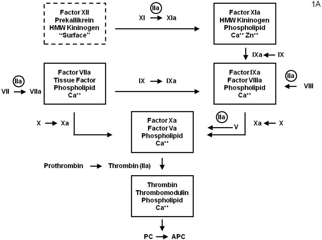
Figure 1A: A diagram illustrating the surface bound complex enzymes of the “intrinsic” and “extrinsic” coagulation pathways. The surface bound complex of the intrinsic pathway is outlined by a dashed line. The amplification by thrombin feedback activations of fV, fVIII, fVII and fXI are also illustrated. Figure 1B: Coagulation system regulation by the stoichiometric (AT and TFPI) inhibitors which down-regulate the system by binding the serine proteases and the dynamic APC system, which causes proteolytic inactivation the cofactors (fVa and fVIIIa). (Figure 1 from Mann KG. Thrombin Formation CHEST 2003; 124:4S–10S with permission)
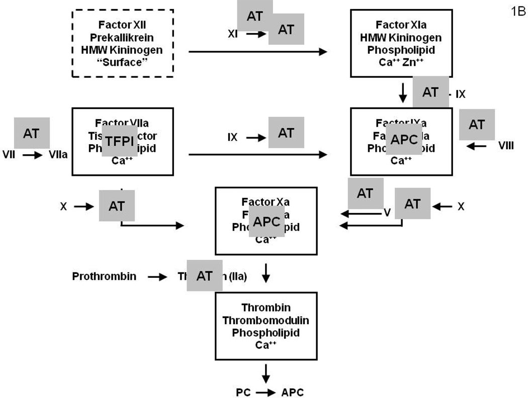
Figure 1A: A diagram illustrating the surface bound complex enzymes of the “intrinsic” and “extrinsic” coagulation pathways. The surface bound complex of the intrinsic pathway is outlined by a dashed line. The amplification by thrombin feedback activations of fV, fVIII, fVII and fXI are also illustrated. Figure 1B: Coagulation system regulation by the stoichiometric (AT and TFPI) inhibitors which down-regulate the system by binding the serine proteases and the dynamic APC system, which causes proteolytic inactivation the cofactors (fVa and fVIIIa). (Figure 1 from Mann KG. Thrombin Formation CHEST 2003; 124:4S–10S with permission)
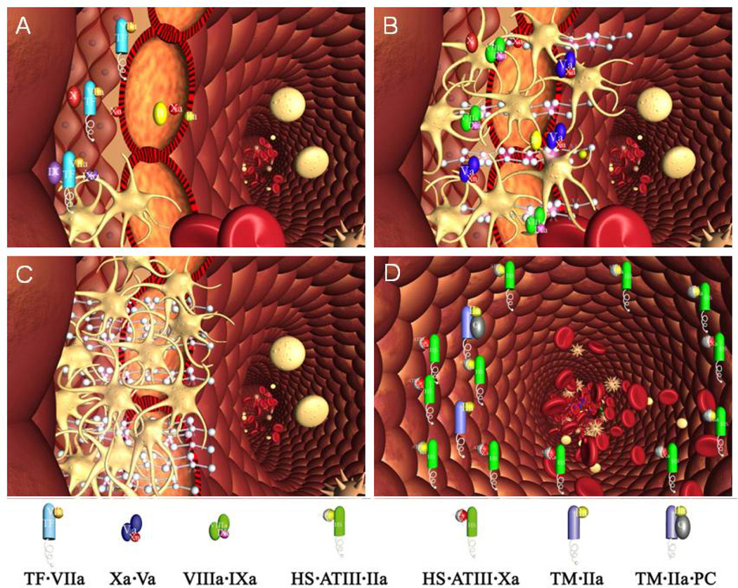
A cross section of a blood vessel showing the luminal space, endothelial cell layer and extravascular region is presented at the site of a perforation. The blood coagulation process in response is depicted in four stages. Tissue factor-factor VIIa complex, TF•VIIa; prothrombinase complex, Xa•Va; intrinsic factor Xase, VIIIa•IXa; ATIII-endothelial cell heparan sulfate proteoglycan complex bound to thrombin or factor Xa, HS•ATIII• (IIa or Xa); protein C bound to thrombomodulin-thrombin,TM•IIa•PC. Panel A. Perforation results in delivery of blood, and with it circulating factor VIIa and platelets, to an extravascular space rich in membrane bound TF. Platelets adhere to collagen and von Willebrand factor associated with the extravascular tissue, and TF binds factor VIIa, initiating the process of factor IX and factor X activation. Factor Xa activates small amounts of prothrombin to thrombin that activates more platelets and converts factor V and factor VIII to factor Va and factor VIIIa. Panel B. The reaction is propagated by platelet-bound intrinsic factor Xase and prothrombinase with the former being the principle factor Xa generator. Initial clotting occurs and fibrin begins to fill in the void in cooperation with activated platelets. Panel C. A barrier composed of activated platelets ladened with procoagualant complexes and enmeshed in fibrin scaffolding is formed. The reaction in the now filled perforation is terminated by reagent consumption attenuating further thrombin generation but functional procoagulant enzyme complexes persist because they are protected from the dynamic inhibitory processes found on the intravascular face. Panel D. View downstream of the perforation. Enzymes escaping from the plugged perforation are captured by antithrombin-heparan complexes and the protein C system is activated by residual thrombin binding to endothelial cell thrombomodulin, initiating the dynamic anticoagulant system. These intravascular processes work against occlusion of the vessel despite the continuous resupply of reactants across the intravascular face of the thrombus. From Orfeo, J. Biol. Chem. 280:42887–42896, 2005. With permission.
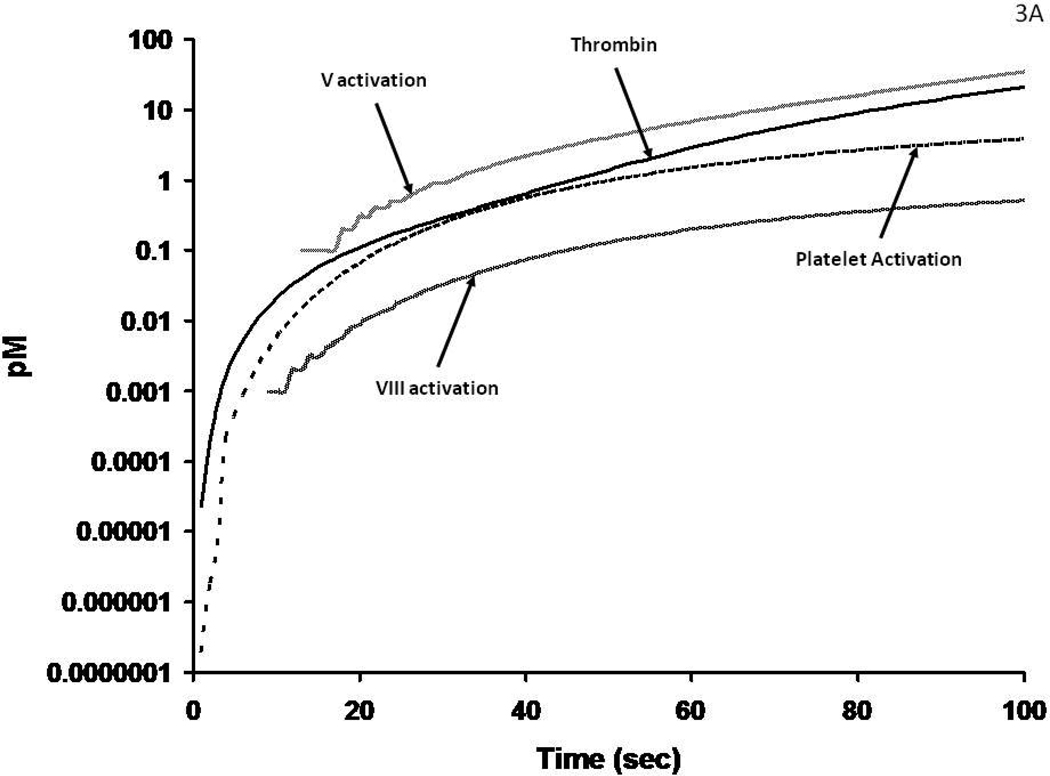
Figure 3A: Numerical simulations of1 the earliest events leading to catalyst formation with a Tf insult. The scale is logarithmic illustrating the initial activation over time of fV, fVIII and platelets by the thrombin initially produced (by fXa) in the reaction. Figure 3B: The generation of thrombin, fVa and fVIIIa and platelet activation over the entire course of the reaction. The thrombin catalyzed feedback amplifications resulting in enhanced platelets and fVIII activation associated with the generation of increased amounts of thrombin by the prothrombinase complex are shown in this numerical simulation.
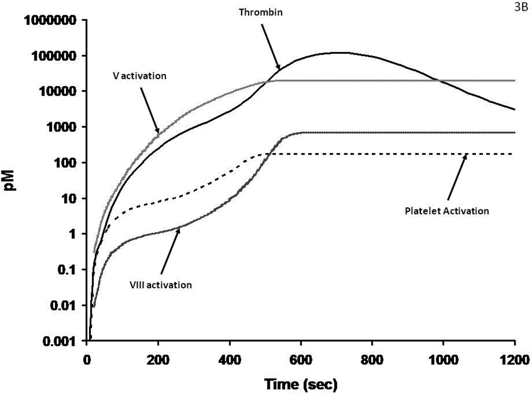
Figure 3A: Numerical simulations of1 the earliest events leading to catalyst formation with a Tf insult. The scale is logarithmic illustrating the initial activation over time of fV, fVIII and platelets by the thrombin initially produced (by fXa) in the reaction. Figure 3B: The generation of thrombin, fVa and fVIIIa and platelet activation over the entire course of the reaction. The thrombin catalyzed feedback amplifications resulting in enhanced platelets and fVIII activation associated with the generation of increased amounts of thrombin by the prothrombinase complex are shown in this numerical simulation.
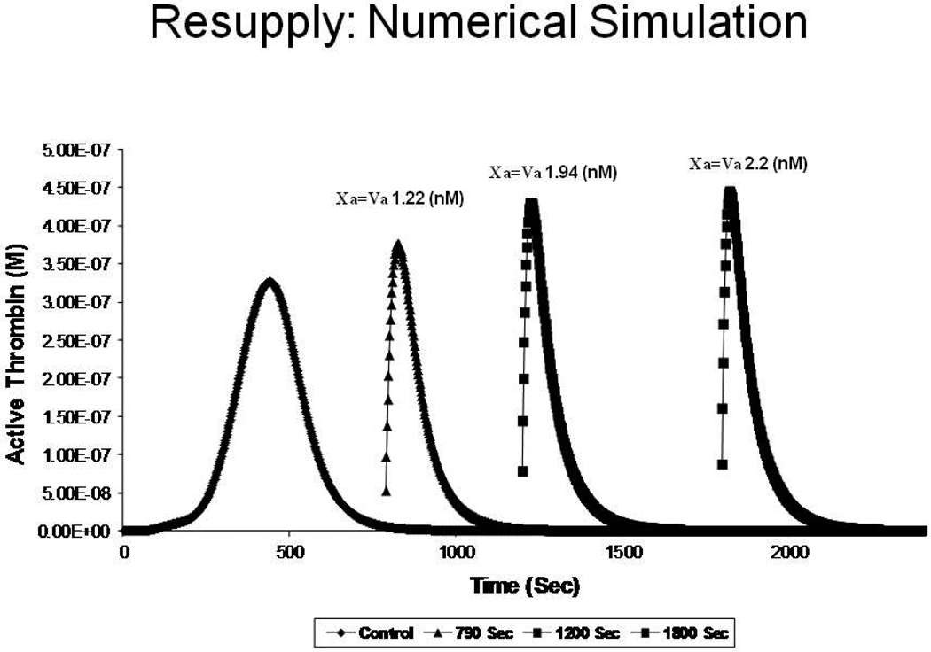
A numerical simulation of consecutive resupply of “electronic” plasma reactants to a quiescent thrombus; at 790, 1200 and 1800 seconds resulting in a more intense thrombin generation and providing higher concentrations of prothrombinase (Xa = Va). From Orfeo, J. Biol. Chem. 280:42887–42896, 2005. With permission.
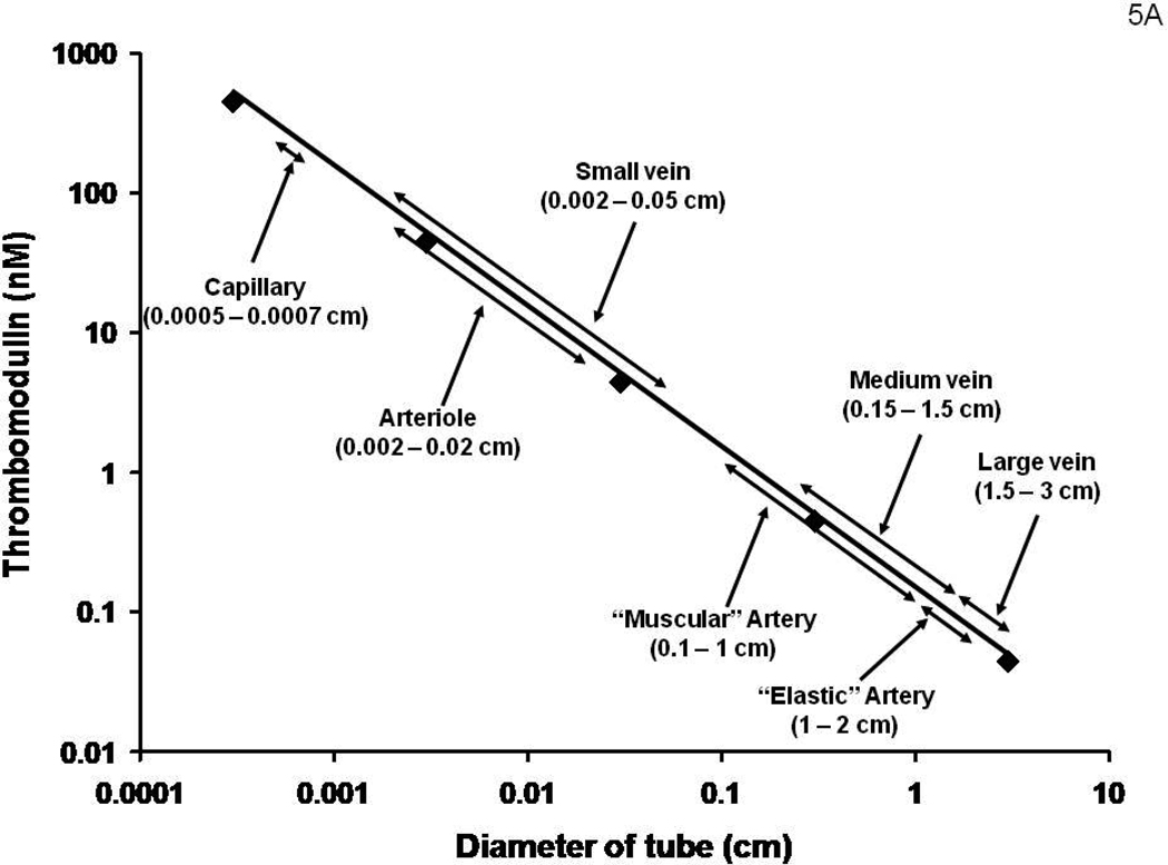
Figure 5A: A hypothetical construct displaying the potential molar concentrations of Tm in various regions of the vasculature. An underlying assumption is that the Tm concentration by each endothelial cell is independent of the vascular source. Figure 5B: A numerical simulation of thrombin generation versus time in vessels displaying the Tm concentrations estimated in Figure 5A following a Tf insult.
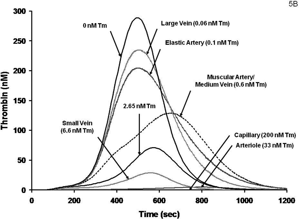
Figure 5A: A hypothetical construct displaying the potential molar concentrations of Tm in various regions of the vasculature. An underlying assumption is that the Tm concentration by each endothelial cell is independent of the vascular source. Figure 5B: A numerical simulation of thrombin generation versus time in vessels displaying the Tm concentrations estimated in Figure 5A following a Tf insult.
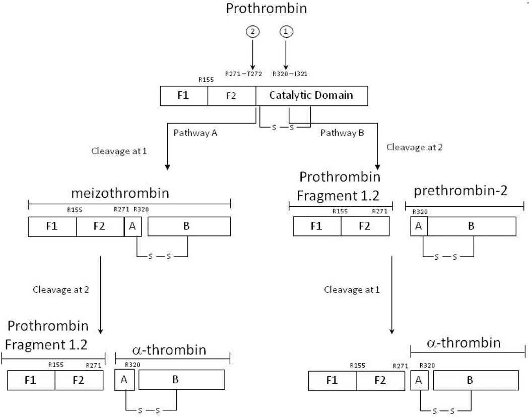
Pathways of Prothrombin Activation. The order of the two bond cleavages yield either the thrombin precursor prethrombin-2 or the enzyme, meizothrombin, as intermediates From Krishnaswamy J. Biol. Chem. 262:3291-99, 1987. With permission.
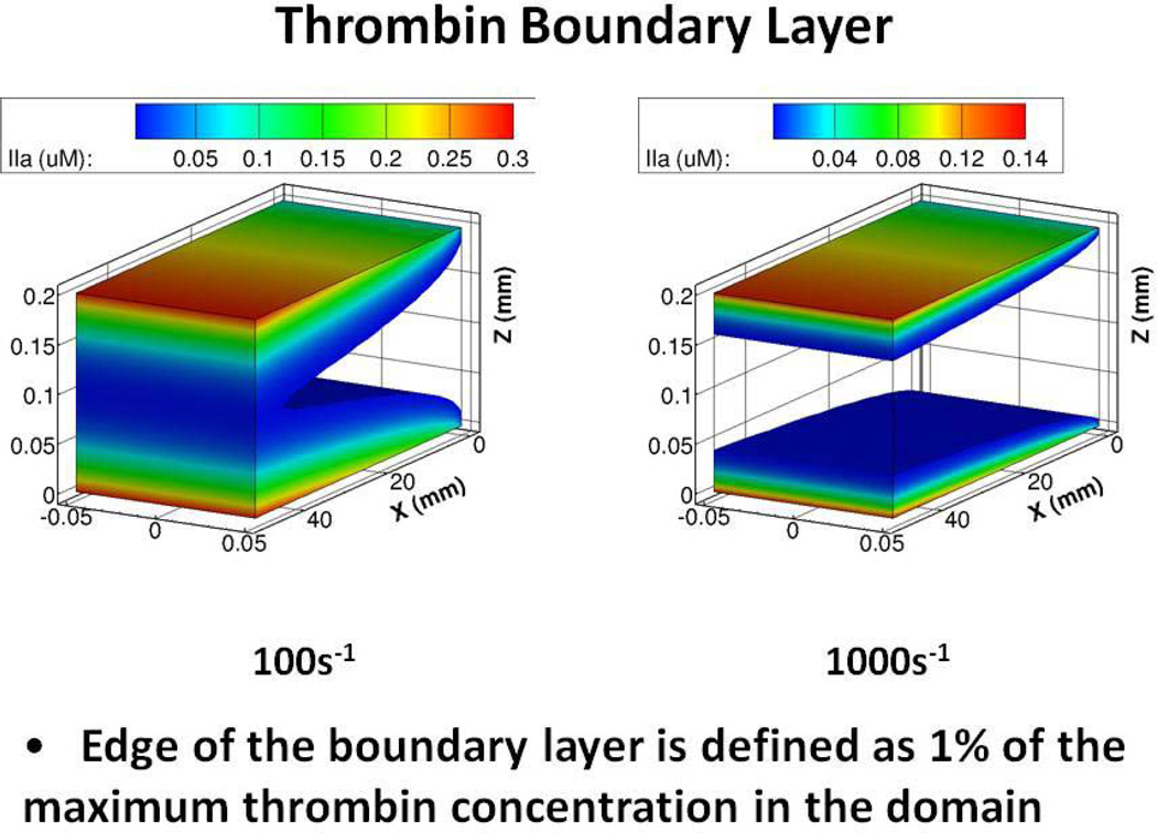
Computational flow dynamic model of thrombin concentrations extending from the wall site of formation at venous (100s−1) and arterial (1000s−1) shear rates. (from the data of Haynes et al (with permission) . Figure provided by Yves Dubief.
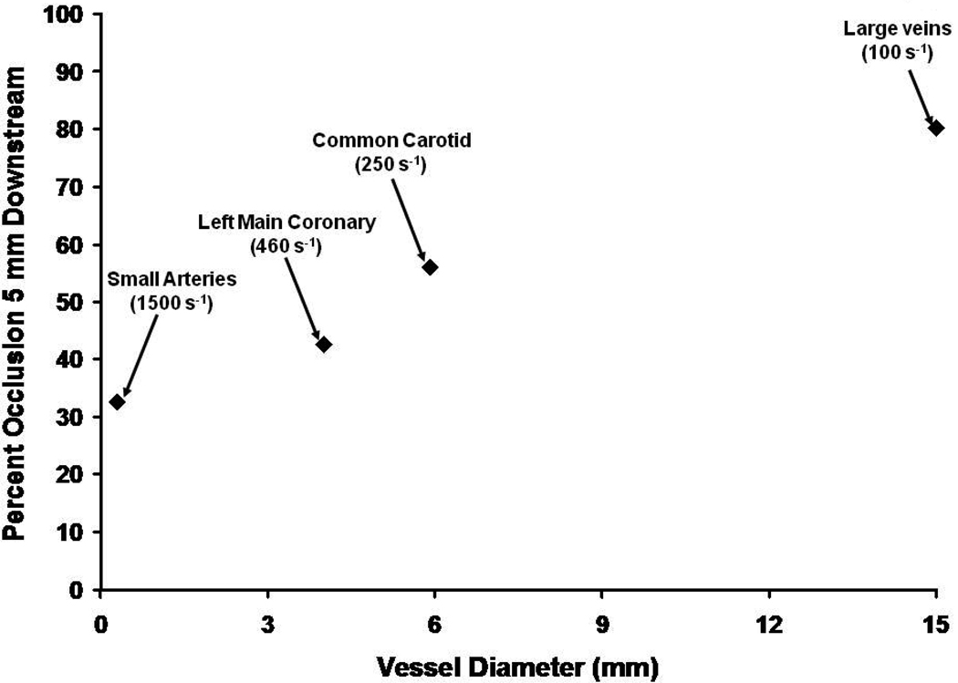
A hypothetical estimate of the extent of vascular occlusion which would occur 5 mm downstream from an anchored thrombin generating catalyst (prothrombinase) in the absence of platelets. The illustration is based upon a combination of fluid mechanics analyses with empirically defined enzyme kinetics studied in a flowing system. The data suggest that in smaller vessels at high shear rates a Tf induced thrombin generation with plasma constituents alone would not lead to occlusion without another source of vascular obstruction (i.e. a platelet thrombus).
References
-
- Owen CA., Jr . A History of Blood Coagulation. Rochester, MN: Mayo Foundation for Medical Education and Research; 2001.
-
- Virchow R. "Thrombose und Embolie. Gefässentzündung und septische Infektion". In: Matzdorff AC, Bell WR, editors. Gesammelte Abhandlungen zur wissenschaftlichen Medicin. Translation. Canton, Massachusetts: Science History Publications; 1998. p. 1856.
-
- Herrick JB. Landmark article (JAMA 1912). Clinical features of sudden obstruction of the coronary arteries. JAMA. 1983;250:1757–1765. - PubMed
-
- DeWood MA, Spores J, Notske R, Mouser LT, Burroughs R, Golden MS, Lang HT. Prevalence of total coronary occlusion during the early hours of transmural myocardial infarction. N.Engl.J.Med. 1980;303:897–902. - PubMed
-
- Hillis LD, Borer J, Braunwald E, Chesebro JH, Cohen LS, Dalen J, Dodge HT, Francis CK, Knatterud G, Ludbrook P, Markis JE, Mueller H, Desvigne-Nickens P, Passamani ER, Powers ER, Rao AK, Roberts R, Roberts WC, Ross A, Ryan TJ, Sobel BE, Williams DO, Zaret BL Co-Investigators. High dose intravenous streptokinase for acute myocardial infarction: preliminary results of a multicenter trial. J Am Coll Cardiol. 1985;6:957–962. - PubMed
Publication types
MeSH terms
Substances
Grants and funding
LinkOut - more resources
Full Text Sources
Other Literature Sources
Medical
