Review
. 2011 Jul 12;77(2):179-89.
doi: 10.1212/WNL.0b013e318224afde.

Encephalitis and antibodies to synaptic and neuronal cell surface proteins

Affiliations
Review

Encephalitis and antibodies to synaptic and neuronal cell surface proteins

Eric Lancaster et al. Neurology. .

Abstract

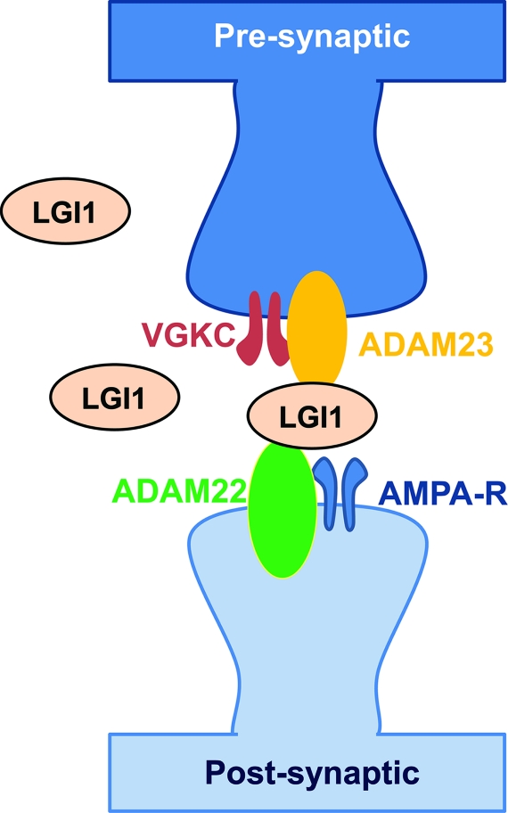
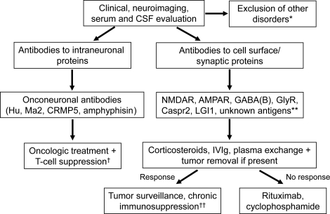
The identification of encephalitis associated with antibodies against cell surface and synaptic proteins, although recent, has already had a substantial impact in clinical neurology and neuroscience. The target antigens are receptors and proteins that have critical roles in synaptic transmission and plasticity, including the NMDA receptor, the AMPA receptor, the GABA(B) receptor, and the glycine receptor. Other autoantigens, such as leucine-rich glioma-inactivated 1 and contactin-associated protein-like 2, form part of trans-synaptic complexes and neuronal cell adhesion molecules involved in fine-tuning synaptic transmission and nerve excitability. Syndromes resulting from these immune responses resemble those of pharmacologic or genetic models in which the antigens are disrupted. For some immune responses, there is evidence that the antibodies alter the structure and function of the antigen, suggesting a direct pathogenic effect. These disorders are important because they can affect children and young adults, are severe and protracted, occur with or without tumor association, and respond to treatment but may relapse. This review provides an update on these syndromes and autoantigens with special emphasis on clinical diagnosis and treatment.
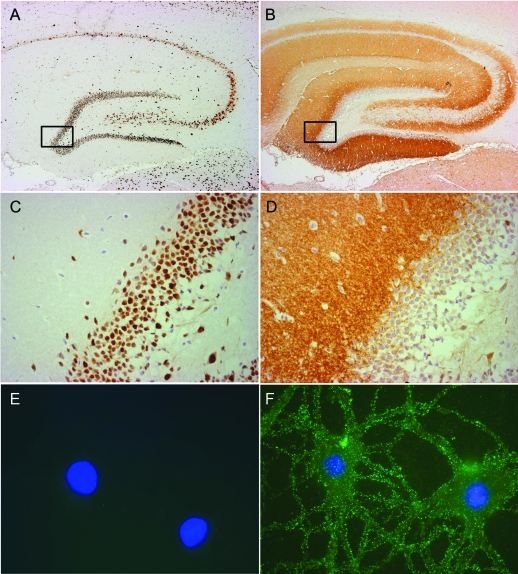
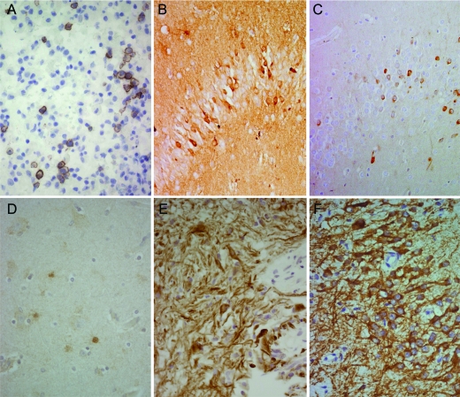
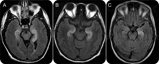

Figures

Figure 1
Figure 1. Comparative analysis of antibodies to intracellular and cell surface antigens
Consecutive sections of rat hippocampus immunostained with CSF of a patient with antibodies against an intracellular antigen (Hu, A) and CSF of a patient with antibodies against a cell surface synaptic antigen (NMDA receptor [NMDAR], B). (C, D) Framed areas shown at higher magnification. Compared with the intracellular antigen, the cell surface synaptic antigen is demonstrated as intense neuropil staining, sparing neuronal cell bodies (nuclei of neurons mildly counterstained with hematoxylin). Using live, nonpermeabilized cultures of dissociated rat hippocampal neurons, the Hu antibody does not show reactivity due to lack of penetration into the neuron (E), whereas the NMDAR antibody shows intense neuronal cell surface immunolabeling (F), indicating that it recognizes an extracellular epitope (nuclei of neurons counterstained in blue with 4′,6-diamidino-2-phenylindole). (A–D) Immunoperoxidase method (A, B 10; C, D 400). (E, F) Immunofluorescence method (800 oil lens). (A–D) Modified, with permission, from Tuzun and Dalmau7 (Limbic encephalitis and variants: classification, diagnosis and treatment. Neurologist 2007;13:261–271).
Figure 2
Figure 2. Presence of plasma cells and immunoglobulin G (IgG) but not complement in the brain of patients with anti-NMDA receptor (NMDAR) encephalitis
Demonstration of plasma cells (A), deposits of IgG (B, diffuse brown staining of neuropil), and absence of complement (D) in the brain from the autopsy of a patient with anti-NMDAR encephalitis. (C) Corresponds to the hippocampus of a neurologically normal, NMDAR antibody–negative individual; note the absence of IgG compared with B. The lack of complement in the patient's brain is in contrast with the detection of complement in the associated ovarian teratoma (E). Cells and processes contained in the tumor react with MAP-2, a neuronal dendritic marker (F). Specific markers used include anti-CD138 (specific for plasma cells, A), anti-human IgG (B, C), anti-C9neo (specific for activated complement, D, E), and anti-MAP-2 (specific for dendrites, F). Immunoperoxidase technique, all panels 400.
Figure 3
Figure 3. MRI of patients with limbic encephalitis and different antibodies to cell surface antigens
The MRI scans correspond to patients with LGI1 antibodies (A), AMPA-R antibodies (B), and GABAB-R antibodies (C).
Figure 4
Figure 4. Interaction of leucine-rich glioma-inactivated 1 (LGI1) with presynaptic and postsynaptic proteins
LGI1 is a secreted neuronal protein that interacts at the presynapse with ADAM23 and at the postsynapse with ADAM22. The cartoon is based on studies indicating that LGI1 coprecipitates with other proteins including the presynaptic Kv1 potassium channels and a variety of presynaptic and postsynaptic scaffolding proteins.,e53 It has been postulated that LGI1 connects presynaptic and postsynaptic protein complexes for finely tuned synaptic transmission. AMPAR = α-amino-3-hydroxy-5-methyl-4-isoxazolepropionic acid receptor.
Figure 5
Figure 5. Algorithmic approach to diagnosis and treatment of encephalitis with antibodies to intracellular and cell surface neuronal antigens
See discussion in text. *Differential diagnosis and exclusion of other disorders has been examined in several reviews.,,e54 **Unknown antigens refer to antigens whose identity has not been established but are visible using patients' antibodies with brain immunohistochemistry and cultures of neurons (as in figure 1, B, D, and F). †T-cell suppression refers to strategies focused on decreasing T-cell activation (rituximab) and cytotoxic T-cell mechanisms (cyclophosphamide, tacrolimus, or cyclosporine). ††As indicated in text, tumor surveillance and chronic immunosuppression should be considered in some disorders (α-amino-3-hydroxy-5-methyl-4-isoxazolepropionic acid receptor [AMPAR], γ-aminobutyric acid-B receptor [GABAB-R], and contactin-associated protein-like 2 [Caspr2]) and subgroups of patients with anti-NMDA receptor (NMDAR) encephalitis with higher risk for relapse (e.g., patients without tumor) or to have an underlying tumor (older than 18 years). GlyR = glycine receptor; IVIg = IV immunoglobulin; LGI1 = leucine-rich glioma-inactivated 1.

Similar articles

Cited by

References

    1. Dalmau J, Tuzun E, Wu HY, et al. Paraneoplastic anti-N-methyl-d-aspartate receptor encephalitis associated with ovarian teratoma. Ann Neurol 2007;61:25–36 - PMC - PubMed
    1. Lai M, Hughes EG, Peng X, et al. AMPA receptor antibodies in limbic encephalitis alter synaptic receptor location. Ann Neurol 2009;65:424–434 - PMC - PubMed
    1. Lancaster E, Lai M, Peng X, et al. Antibodies to the GABAB receptor in limbic encephalitis with seizures: case series and characterisation of the antigen. Lancet Neurol 2010;9:67–76 - PMC - PubMed
    1. Lai M, Huijbers MG, Lancaster E, et al. Investigation of LGI1 as the antigen in limbic encephalitis previously attributed to potassium channels: a case series. Lancet Neurol 2010;9:776–785 - PMC - PubMed
    1. Hutchinson M, Waters P, McHugh J, et al. Progressive encephalomyelitis, rigidity, and myoclonus: a novel glycine receptor antibody. Neurology 2008;71:1291–1292 - PubMed

Publication types