. 2011 Sep;52(9):1660-71.
doi: 10.1194/jlr.M017582. Epub 2011 Jul 11.

Unique sphingomyelin patches are targets of a beta-cell-specific antibody

Affiliations

Unique sphingomyelin patches are targets of a beta-cell-specific antibody

Amol Kavishwar et al. J Lipid Res. 2011 Sep.

Abstract

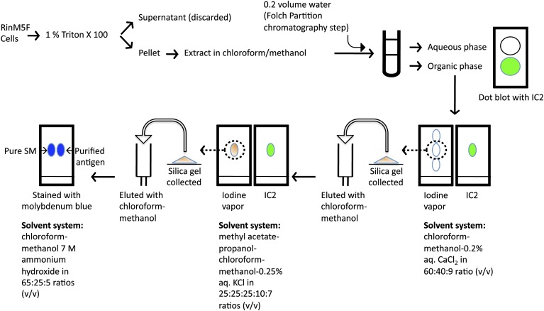
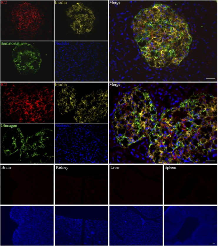
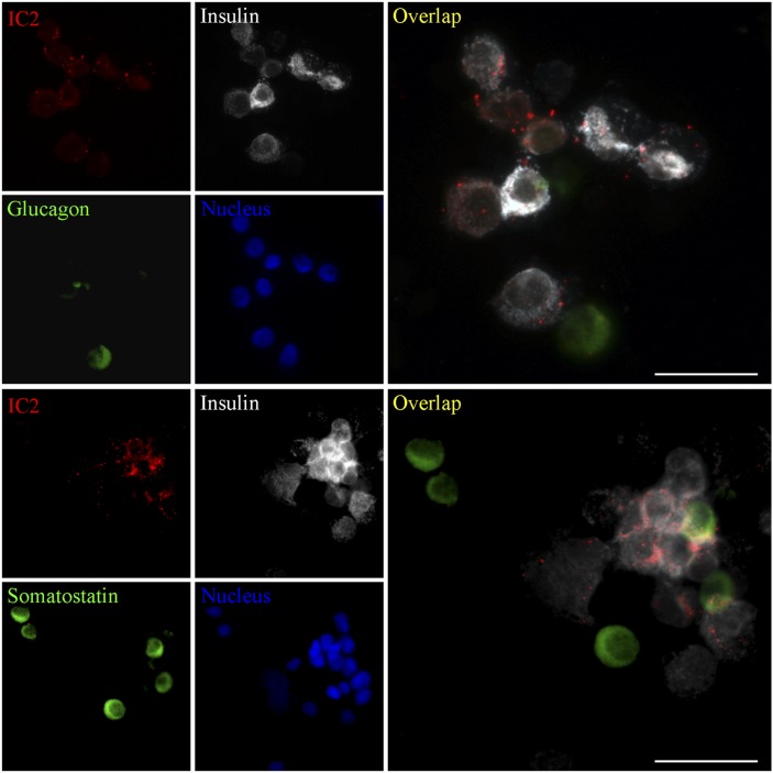
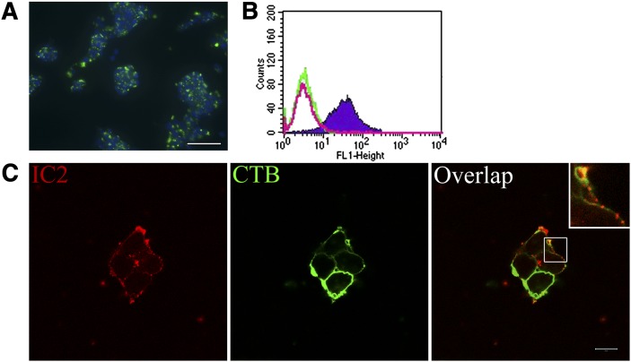
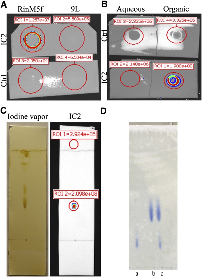
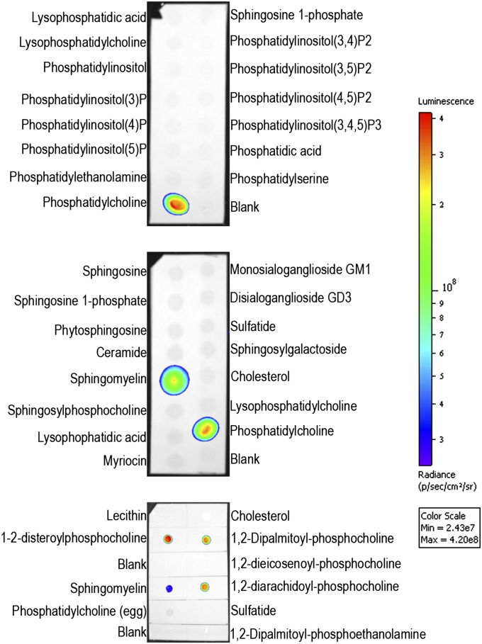
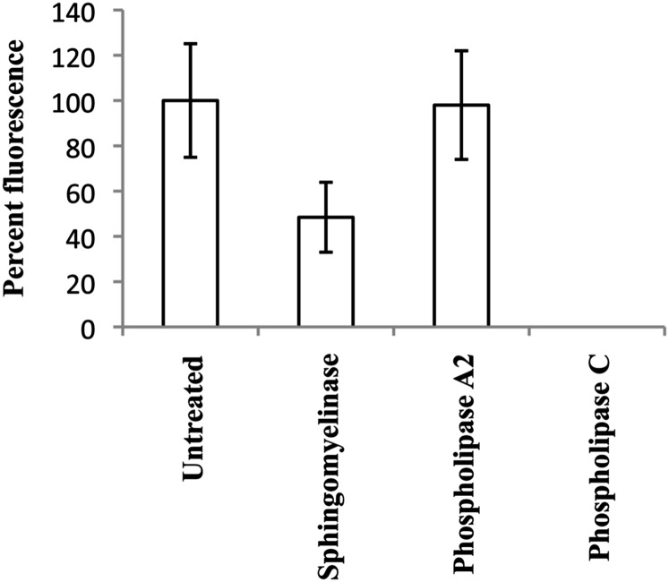
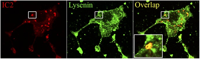
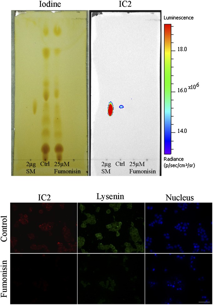
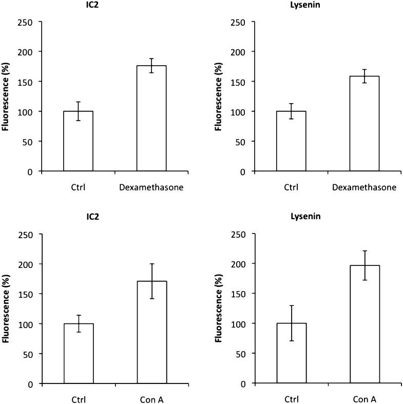
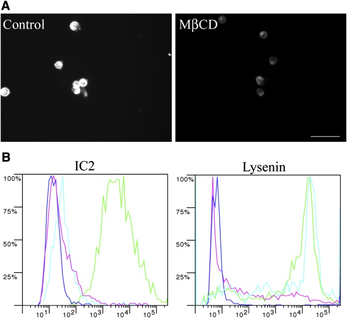
To devise successful imaging and therapeutic strategies, the identification of β-cell surface markers is one of the challenges in diabetes research that has to be resolved. We previously showed that IC2, a rat monoclonal IgM antibody, can be used for ex vivo determination of β-cell mass by imaging. Further progress toward the development of an antibody-based imaging agent was hampered by the lack of knowledge regarding the nature and composition of the IC2 antigen. Here, we show a series of systematic experiments involving classical lipid extraction and chromatography techniques combined with immunochemistry, which led to the identification of sphingomyelin as the target antigen assembled in the form of patches on the β-cell surface. Our findings were verified by modulating SM by enzymatic cleavage, downregulation, upregulation, and perturbation of membrane SM and observation of corresponding changes in IC2 binding. Cholesterol participates in stabilization of these patches, as its removal results in loss of IC2 binding. We believe that these findings have implications for identifying future ligands for the proposed antigen for imaging purposes as well as for potential therapy, as sphingomyelin has been shown to play a role in the apoptotic cascade in pancreatic β cells.


Figures

Fig. 1.
Fig. 1.
Antigen purification scheme. Triton X100 insoluble fraction of RinM5f cells was extracted in methanol-chloroform mixture and then further fractionated and purified following classical chromatography steps. Enrichment of antigen at every step was followed by immunoblotting with IC2 antibody on TLC plates.
Fig. 2.
Fig. 2.
Immunostaining with IC2. Cryosections of human pancreas were stained with IC2 along with insulin and somatostatin or glucagon and observed under a fluorescence microscope. Similarly, cryosections of mouse brain, kidney, liver, and spleen were stained with IC2 alone and observed under a fluorescence microscope. Bar = 100 µm.
Fig. 3.
Fig. 3.
IC2 binds with islet β cells. Human islets were dissociated with trypsin and stained with IC2 along with insulin and glucagon or somatostatin. Bar = 50 µm.
Fig. 4.
Fig. 4.
Cell surface localization of IC2. A: Characteristic punctate staining of RinM5f insulinoma cells shows strong binding with IC2 antibody (IC2, green; DAPI nuclear stain, blue). B: FACS analysis of Ins-1E cells shows specific binding with IC2 antibody (shaded region). No nonspecific uptake was observed with isotype control antibody (green line); unstained cells, red line. C: Confocal microscopy with RinM5f cells confirmed cell surface localization of the antigen. Staining for IC2 (red) colocalized with the staining for membrane marker cholera toxin B subunit (green). Note punctate staining on the membrane (insert). Bar = 10 µm.
Fig. 5.
Fig. 5.
Purification of IC2 antigen. A: Dot blot of chloroform/methanol (1:1) extract of Triton X100-insoluble fraction of RinM5f and 9L cells probed with IC2 antibody. B: The chloroform/methanol extract from RinM5f cells was partitioned into aqueous and organic phase. Dot blot with IC2 antibody demonstrated positive signal in organic phase. C: Lipids present in organic phase were resolved on TLC plate and stained with iodine vapors. After documenting the image, the TLC plate was immunostained with IC2 antibody. D: Purified IC2 antigen (b) was run along with PC (a) and a mixture of SM and PC (c) and stained with molybdenum blue.
Fig. 6.
Fig. 6.
Immunoblotting on lipid array. Two commercially available (PIPStrip and SphingoStrip) and one homemade lipid arrays (bottom) were immunoblotted against IC2 antibody.
Fig. 7.
Fig. 7.
Lipase treatment affects IC2 binding. RinM5f cells were treated with lipases and then stained with IC2. There was a significant decrease in IC2 binding after the treatment with sphingomyelinase and phospholipase C.
Fig. 8.
Fig. 8.
Coimmunolocalization of IC2 and lysenin. RinM5f cells were stained with IC2 and lysenin and observed under a confocal microscope. Bar = 10 µm.
Fig. 9.
Fig. 9.
Fumonisin B1 treatment. Top: Cells were treated with Fumonisin B1, total lipids were extracted, resolved on TLC plate, stained with iodine vapors, and then subjected to immunoblotting against IC2 antibody. Untreated cells (Ctrl) and purified SM served as controls. Bottom: Cells treated with Fumonisin B1 were coimmunostained with IC2 and lysenin and observed under a fluorescence microscope. Bar = 50 µm.
Fig. 10.
Fig. 10.
Upregulation of sphingomyelin. RinM5f cells were treated with dexamethasone or concanavalin A and then stained with IC2 or lysenin. Both treatment regimens resulted in increased signal from the cells.
Fig. 11.
Fig. 11.
MβCD treatment. A: RinM5f cells were treated with MβCD to remove membrane cholesterol and then stained with the cholesterol-specific fluorescent antibiotic Filipin III. Untreated cells served as a positive control. Bar = 50 µm. B: MβCD-treated cells were stained with IC2 or lysenin and analyzed by flow cytometry. Positive control, green; negative control, blue; SMase treatment as the second negative control, red; MβCD-treated cells, cyan.

Similar articles

Cited by

References

    1. Moore A. 2009. Advances in beta-cell imaging. Eur. J. Radiol. 70: 254–257. - PMC - PubMed
    1. Moore A., Bonner-Weir S., Weissleder R. 2001. Noninvasive in vivo measurement of beta-cell mass in mouse model of diabetes. Diabetes. 50: 2231–2236. - PubMed
    1. Antkowiak P., Tersey S., Carter J., Vandsburger M., Nadler J., Epstein F., Mirmira R. 2008. Noninvasive assessment of pancreatic beta-cell function in vivo with manganese-enhanced magnetic resonance imaging. Am. J. Physiol. Endocrinol. Metab. 296: 573–578. - PMC - PubMed
    1. Souza F., Simpson N., Raffo A., Saxena C., Maffei A., Hardy M., Kilbourn M., Goland R., Leibel R., Mann J., et al. 2006. Longitudinal noninvasive PET-based β cell mass estimates in a spontaneous diabetes rat model. J. Clin. Invest. 116: 1506–1513. - PMC - PubMed
    1. Kung M., Hou C., Lieberman B., Oya S., Ponde D., Blankemeyer E., Skovronsky D., Kilbourn M. R., Kung H. 2008. In vivo imaging of β-cell mass in rats using 18F-FP-(+)-DTBZ: a potential PET ligand for studying diabetes mellitus. J. Nucl. Med. 49: 1171–1176. - PubMed

Publication types

MeSH terms

LinkOut - more resources