

Trefoil factor 1 stimulates both pancreatic cancer and stellate cells and increases metastasis
- PMID: 21747314
- PMCID: PMC4319540
- DOI: 10.1097/MPA.0b013e31821f6927
Trefoil factor 1 stimulates both pancreatic cancer and stellate cells and increases metastasis
Abstract
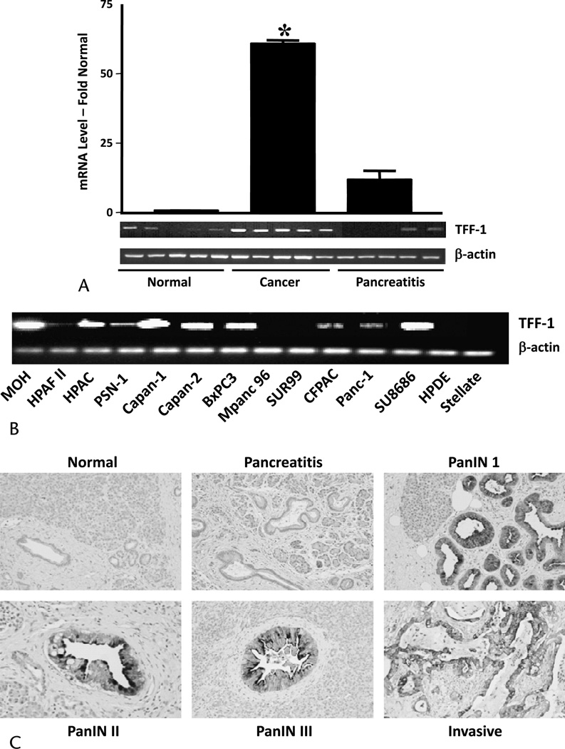
Objectives: Trefoil factor 1 (TFF1) is a stable secretory protein expressed widely in the gastrointestinal mucosa that is also expressed in pancreatic ductal adenocarcinoma (PDAC). In the current study, we documented the extent and timing of TFF1 expression and investigated the effects of TFF1 on PDAC cells and stellate cells, the primary cells of the PDAC stroma.
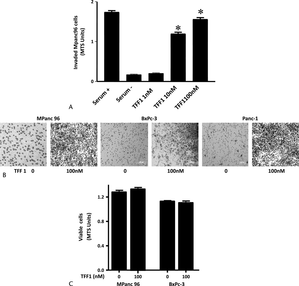
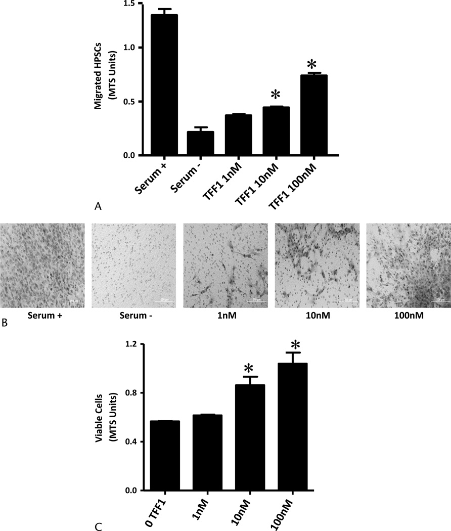
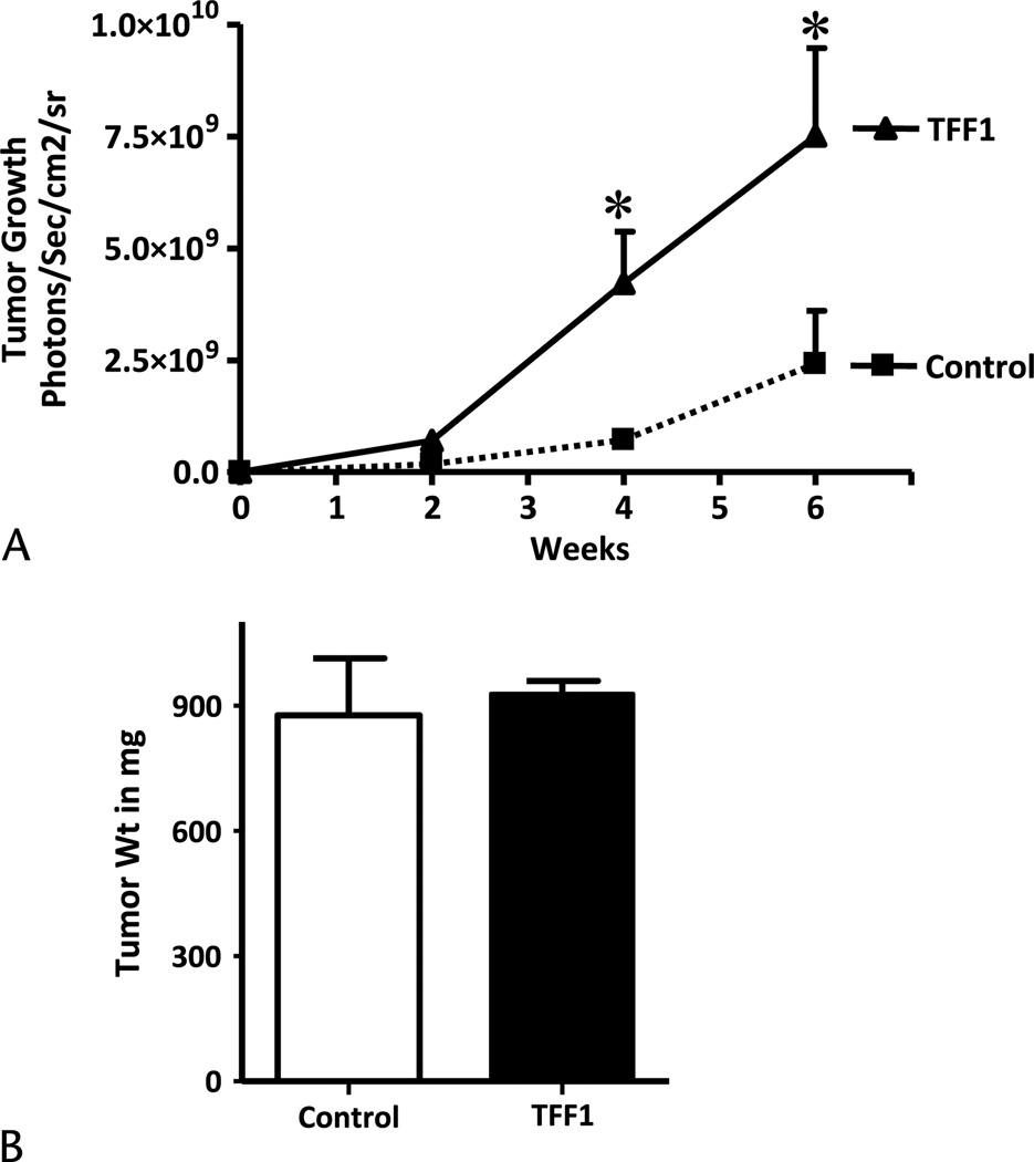
Methods: Trefoil factor 1 expression in pancreatic cancer tissues and cell lines was analyzed using microarray, quantitative reverse transcriptase-polymerase chain reaction, and immunohistochemistry. The effects of recombinant TFF1 on cell growth, migration, and invasion of pancreatic cancer cell lines and immortalized human pancreatic stellate cells (HPSCs) were analyzed using MTS and Matrigel-coated invasion chambers. In vivo studies were also conducted in which Mpanc-96 cells stably expressing TFF1 were implanted orthotopically into nude mice.
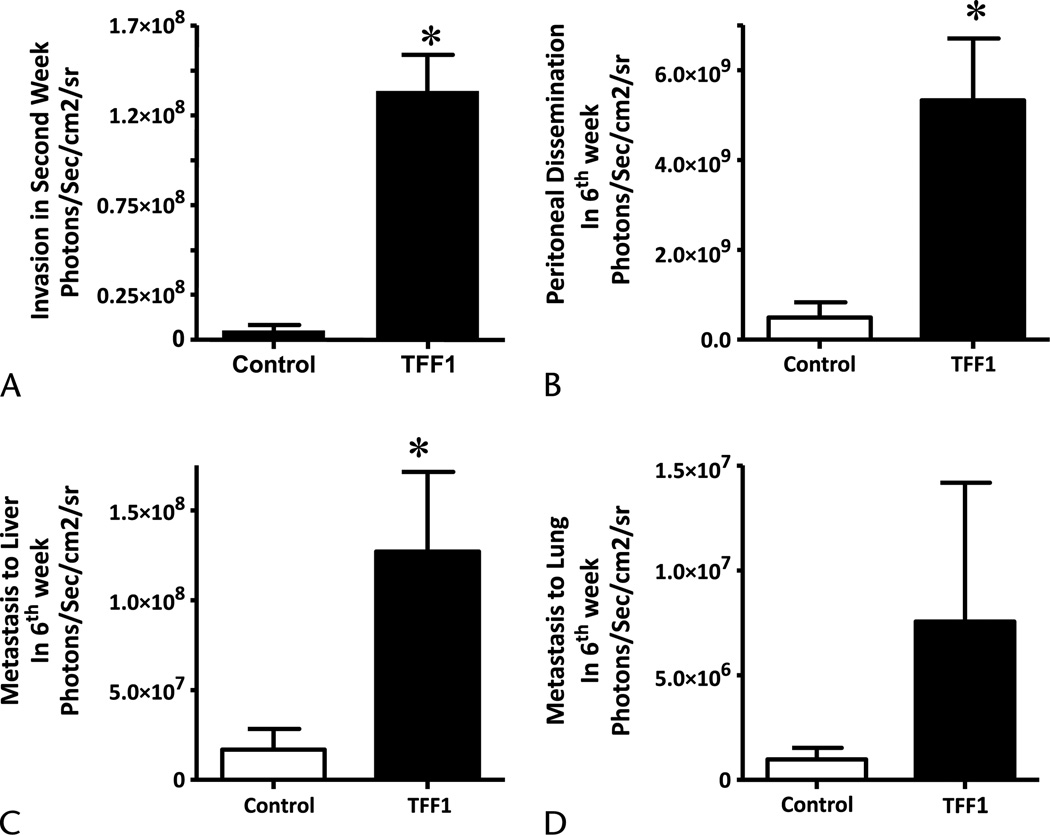
Results: Trefoil factor 1 was highly increased in preneoplastic lesions. Recombinant TFF1 stimulated motility of both cancer and HPSCs. In contrast, only HPSC cell growth was increased by TFF1. In vivo studies showed that overexpression of TFF1 in PDAC cells did not affect primary tumor growth but greatly increased metastasis.
Conclusions: The present data demonstrate that TFF1 influences both PDAC cells and stellate cells and stimulates metastasis.
Conflict of interest statement
The authors have no any potential conflict of interest including any financial, personal, or other relationships with other people or organizations that could inappropriately influence this work.
Figures
Similar articles
-
Secretory Trefoil Factor 1 (TFF1) promotes gemcitabine resistance through chemokine receptor CXCR4 in Pancreatic Ductal Adenocarcinoma.Cancer Lett. 2024 Aug 28;598:217097. doi: 10.1016/j.canlet.2024.217097. Epub 2024 Jul 2. Cancer Lett. 2024. PMID: 38964729
-
Small Nucleolar Noncoding RNA SNORA23, Up-Regulated in Human Pancreatic Ductal Adenocarcinoma, Regulates Expression of Spectrin Repeat-Containing Nuclear Envelope 2 to Promote Growth and Metastasis of Xenograft Tumors in Mice.Gastroenterology. 2017 Jul;153(1):292-306.e2. doi: 10.1053/j.gastro.2017.03.050. Epub 2017 Apr 5. Gastroenterology. 2017. PMID: 28390868
-
Galectin-3 Mediates Tumor Cell-Stroma Interactions by Activating Pancreatic Stellate Cells to Produce Cytokines via Integrin Signaling.Gastroenterology. 2018 Apr;154(5):1524-1537.e6. doi: 10.1053/j.gastro.2017.12.014. Epub 2017 Dec 21. Gastroenterology. 2018. PMID: 29274868
-
Pancreatic stellate cells and pancreas cancer: current perspectives and future strategies.Eur J Cancer. 2014 Oct;50(15):2570-82. doi: 10.1016/j.ejca.2014.06.021. Epub 2014 Aug 1. Eur J Cancer. 2014. PMID: 25091797 Review.
-
Pancreatic stellate cell: Pandora's box for pancreatic disease biology.World J Gastroenterol. 2017 Jan 21;23(3):382-405. doi: 10.3748/wjg.v23.i3.382. World J Gastroenterol. 2017. PMID: 28210075 Free PMC article. Review.
Cited by
-
The pancreatic cancer microenvironment: A true double agent.J Surg Oncol. 2017 Jul;116(1):7-15. doi: 10.1002/jso.24643. Epub 2017 Jun 12. J Surg Oncol. 2017. PMID: 28605029 Free PMC article. Review.
-
Generation and characterization of CRISPR/Cas9-mediated MEN1 knockout BON1 cells: a human pancreatic neuroendocrine cell line.Sci Rep. 2020 Sep 3;10(1):14572. doi: 10.1038/s41598-020-71516-7. Sci Rep. 2020. PMID: 32884006 Free PMC article.
-
Channeling the Force: Piezo1 Mechanotransduction in Cancer Metastasis.Cells. 2021 Oct 20;10(11):2815. doi: 10.3390/cells10112815. Cells. 2021. PMID: 34831037 Free PMC article. Review.
-
Gastric Proteins MUC5AC and TFF1 as Potential Diagnostic Markers of Colonic Sessile Serrated Adenomas/Polyps.Am J Clin Pathol. 2016 Nov 1;146(5):530-537. doi: 10.1093/ajcp/aqw142. Am J Clin Pathol. 2016. PMID: 28430953 Free PMC article.
-
The influence of matrix properties on growth and morphogenesis of human pancreatic ductal epithelial cells in 3D.Biomaterials. 2013 Jul;34(21):5117-27. doi: 10.1016/j.biomaterials.2013.03.086. Epub 2013 Apr 19. Biomaterials. 2013. PMID: 23602364 Free PMC article.
References
-
- Jemal A, Siegel R, Ward E, et al. Cancer statistics. CA Cancer J Clin. 2006;56:106–130. - PubMed
-
- Parkin DM, Bray FI, Devesa SS. Cancer burden in the year 2000. The global picture. Eur J Cancer. 2001;37:S4–S66. - PubMed
-
- Liotta LA, Kohn EC. The microenvironment of the tumour-host interface. Nature. 2001;411:375–379. - PubMed
-
- Fidler IJ. The organ microenvironment and cancer metastasis. Differentiation. 2002;70:498–505. - PubMed
Publication types
MeSH terms
Substances
Grants and funding
LinkOut - more resources
Full Text Sources
Other Literature Sources
Medical
Research Materials
