A clinical and histopathological study of 122 cases of dermatofibroma (benign fibrous histiocytoma)
- PMID: 21747617
- PMCID: PMC3130861
- DOI: 10.5021/ad.2011.23.2.185
A clinical and histopathological study of 122 cases of dermatofibroma (benign fibrous histiocytoma)
Abstract
Background: Many variants of dermatofibromas have been described, and being aware of the variants of dermatofibromas is important to avoid misdiagnosis.
Objective: We wanted to evaluate the clinical and pathologic characteristics of 122 cases of dermatofibromas.
Methods: We retrospectively reviewed the medical records and 122 biopsy specimens of 92 patients who were diagnosed with dermatofibroma in the Department of Dermatology at Eulji Hospital of Eulji University between January 2000 and March 2010.
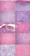
Results: Nearly 80% of the cases occurred between the ages of 20 and 49 years, with an overall predominance of females. Over 70% of the lesions were found on the extremities. The most common histologic variant was a fibrocollagenous dermatofibroma (40.1%). Other variants included histiocytic (13.1%), cellular (11.5%), aneurysmal (7.4%), angiomatous (6.5%), sclerotic (6.5%), monster (4.9%), palisading (1.6%) and keloidal dermatofibromas (0.8%). There were 9 dermatofibromas (7.3%) that were the mixed type with two co-dominant histologic features.
Conclusion: The results of this study are consistent with previous reports on the clinical features of dermatofibromas. However, we observed several characteristic subtypes of dermatofibroma and we compared the frequency of the histologic subtypes.
Keywords: Dermatofibroma.
Figures


Comment in
-
The diagnostic significance of infiltration pattern and perilesional lymphoid cell infiltrate in dermatofibroma.Ann Dermatol. 2012 May;24(2):228-9. doi: 10.5021/ad.2012.24.2.228. Epub 2012 Apr 26. Ann Dermatol. 2012. PMID: 22577281 Free PMC article. No abstract available.
References
-
- Rahbari H, Mehregan AH. Adnexal displacement and regression in association with histiocytoma (dermatofibroma) J Cutan Pathol. 1985;12:94–102. - PubMed
-
- Requena L, Fariña MC, Fuente C, Piqué E, Olivares M, Martín L, et al. Giant dermatofibroma. A little-known clinical variant of dermatofibroma. J Am Acad Dermatol. 1994;30:714–718. - PubMed
-
- De Hertogh G, Bergmans G, Molderez C, Sciot R. Cutaneous cellular fibrous histiocytoma metastasizing to the lungs. Histopathology. 2002;41:85–86. - PubMed
-
- Niemi KM. The benign fibrohistiocytic tumours of the skin. Acta Derm Venereol Suppl (Stockh) 1970;50(Suppl. 63):1–66. - PubMed
-
- Iwata J, Fletcher CD. Lipidized fibrous histiocytoma: clinicopathologic analysis of 22 cases. Am J Dermatopathol. 2000;22:126–134. - PubMed
LinkOut - more resources
Full Text Sources

