A case of an unusual eccrine poroma on the left forearm area
- PMID: 21747633
- PMCID: PMC3130877
- DOI: 10.5021/ad.2011.23.2.250
A case of an unusual eccrine poroma on the left forearm area
Abstract
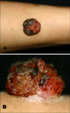
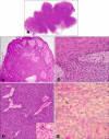
A 40-year-old woman presented with an asymptomatic red to brown colored walnut-sized, dome shaped, hemorrhagic, crusted nodule on the left forearm. There was no previous history of trauma to the area. The first impression of this case was a vascular tumor or malignant lesion due to the large size and bleeding tendency. However, the final diagnosis, according to histologic and immunostaining methods, was a benign eccrine poroma that occurred on the left forearm, which is an unusual area for such a lesion. The tumor was excised and no recurrence was noted when she was examined 24 months later.
Keywords: Eccrine poroma; Forearm.
Figures


References
-
- Goldman P, Pinkus H, Rogin JR. Eccrine poroma; tumors exhibiting features of the epidermal sweat duct unit. AMA Arch Derm. 1956;74:511–521. - PubMed
-
- LoBuono P, Kahn R, Kornblee LV. Eccrine poroma of the forehead. Mt Sinai J Med. 1977;44:527–529. - PubMed
-
- Jin WW, Jung JG, Ro KW, Shim SD, Kim MH, Cinn YW. A case of eccrine poroma on the paranasal area. Ann Dermatol. 2006;18:73–76.
-
- Park SJ, Roh BH, Lee JS, Cho MK, Whang KU. A case of eccrine poroma on the scalp. Korean J Dermatol. 2006;44:633–635.
-
- Johnson RC, Rosenmeier GJ, Keeling JH., 3rd A painful step. Eccrine poroma. Arch Dermatol. 1992;128:1530. - PubMed
Publication types
LinkOut - more resources
Full Text Sources

