. 2011:2011:420905.
doi: 10.1155/2011/420905. Epub 2011 May 29.

Chlamydia trachomatis immune evasion via downregulation of MHC class I surface expression involves direct and indirect mechanisms

Affiliations

Chlamydia trachomatis immune evasion via downregulation of MHC class I surface expression involves direct and indirect mechanisms

Joyce A Ibana et al. Infect Dis Obstet Gynecol. 2011.

Abstract

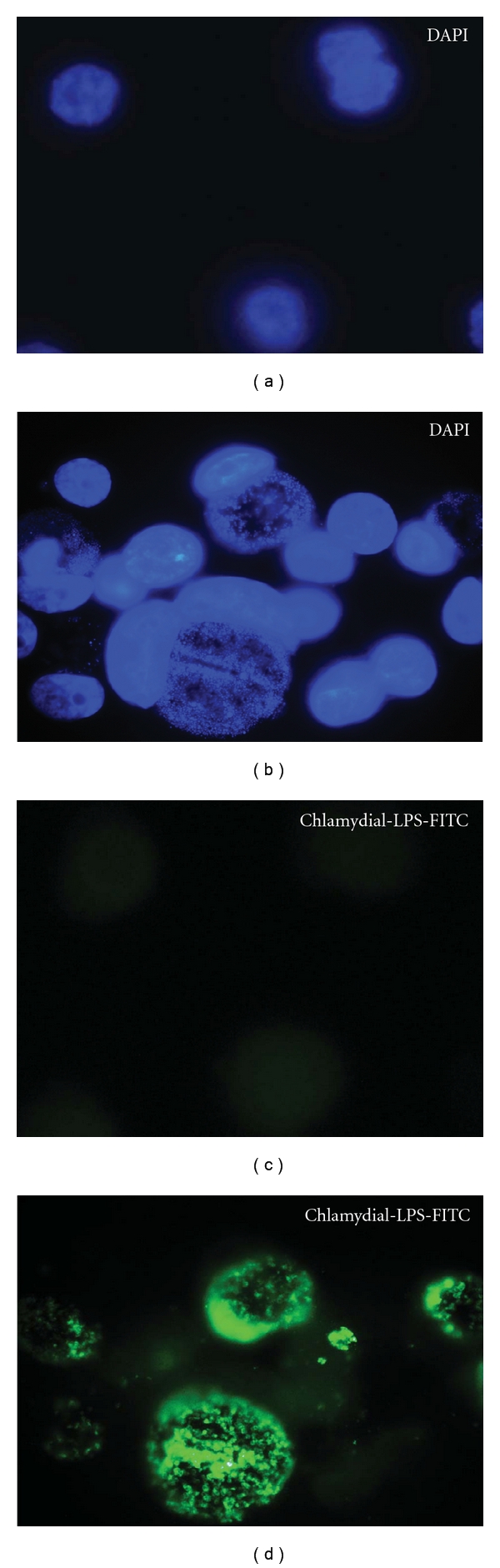
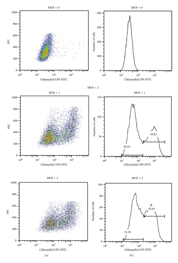
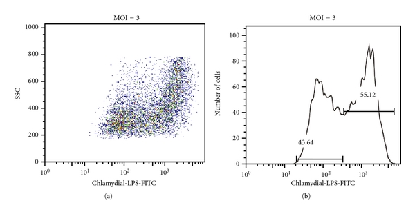
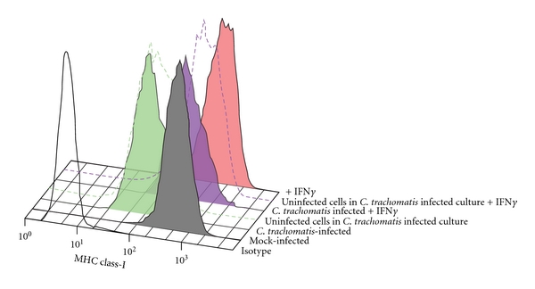
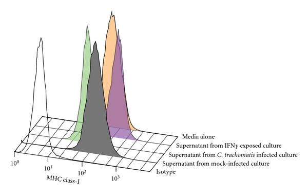
Genital C. trachomatis infections typically last for many months in women. This has been attributed to several strategies by which C. trachomatis evades immune detection, including well-described methods by which C. trachomatis decreases the cell surface expression of the antigen presenting molecules major histocompatibility complex (MHC) class I, MHC class II, and CD1d in infected genital epithelial cells. We have harnessed new methods that allow for separate evaluation of infected and uninfected cells within a mixed population of chlamydia-infected endocervical epithelial cells to demonstrate that MHC class I downregulation in the presence of C. trachomatis is mediated by direct and indirect (soluble) factors. Such indirect mechanisms may aid in priming surrounding cells for more rapid immune evasion upon pathogen entry and help promote unfettered spread of C. trachomatis genital infections.


Figures

Figure 1
Figure 1
C. trachomatis-infected endocervical epithelial cell (A2En) culture. The immortalized endocervical epithelial cell line A2EN was exposed to C. trachomatis serovar D at multiplicity of infection of 2 in the absence of cyclohexamide (right two panels). This resulted in a mixed population of infected and uninfected cells. Infected cells are stained green with chlamydial LPS conjugated to FITC, and nucleic acid is stained with DAPI (blue). Uninfected controls are included in the left two panels for comparison. Data is representative of experiments performed greater than 10 times.
Figure 2
Figure 2
Separation of infected and uninfected endocervical epithelial cells in a C. trachomatis-exposed culture. Using chlamydial-LPS fluorescently labeled with FITC, infected and noninfected A2EN cell populations from the same culture can be distinguished and analyzed independently. This separation allows for the analysis of specific host cellular responses to C. trachomatis infection in infected and in bystander-uninfected cells. Left column shows side scatter (SSC) and chlamydial LPS-FITC dot plots, indicating a shift in granularity of FITC positive cells. Right column shows cell number and chlamydial-LPS histograms, which reflect the proportion of uninfected and C. trachomatis-infected cells within a pool of cells from A2En cultures exposed to varying multiplicities of infection (MOIs). Data is representative of experiments performed greater than 10 times.
Figure 2
Figure 2
Separation of infected and uninfected endocervical epithelial cells in a C. trachomatis-exposed culture. Using chlamydial-LPS fluorescently labeled with FITC, infected and noninfected A2EN cell populations from the same culture can be distinguished and analyzed independently. This separation allows for the analysis of specific host cellular responses to C. trachomatis infection in infected and in bystander-uninfected cells. Left column shows side scatter (SSC) and chlamydial LPS-FITC dot plots, indicating a shift in granularity of FITC positive cells. Right column shows cell number and chlamydial-LPS histograms, which reflect the proportion of uninfected and C. trachomatis-infected cells within a pool of cells from A2En cultures exposed to varying multiplicities of infection (MOIs). Data is representative of experiments performed greater than 10 times.
Figure 3
Figure 3
MHC class I surface expression on infected and uninfected bystander endocervical epithelial cells in cultures exposed to C. trachomatis. Surface expression levels of MHC class I were assessed in the two populations of cells found in C. trachomatis-exposed cultures. A2En cells were either mock infected (gray) or exposed to C. trachomatis at MOI of 2, with cKSFM alone (green) or cKSFM with 30 ng/mL IFN gamma (blue). Mock-infected A2EN cells exposed to 30 ng/mL IFN gamma (red) were analyzed in parallel. Isotype control is shown as black line histogram. Surface expression of MHC class I is depicted using histograms. The green solid histogram represents C. trachomatis-infected cells, the dashed green line represents bystander uninfected cells, the blue solid histogram represents C. trachomatis-infected cells exposed to IFN gamma, and the blue dashed line represents uninfected cells in IFN gamma-exposed cultures. Downregulation of MHC class I expression was observed on both the C. trachomatis-infected cells and on bystander cells. Data is representative of experiments performed in duplicate three times.
Figure 4
Figure 4
Effect of C. trachomatis-exposed A2EN cell culture media on MHC class I surface expression on uninfected A2EN endocervical epithelial cells. Uninfected monolayers of A2EN cells were exposed to culture media from C. trachomatis-infected A2EN cell culture (green), mock-infected culture (gray), IFN gamma exposed infected cell culture (blue), and fresh culture media (red). An isotype control is shown as black line histogram. Exposure of endocervical cells to culture media from C. trachomatis-infected culture at an MOI of 2 for 38 hours, (during which ~50% of cells are infected with C. trachomatis) resulted in the downregulation of MHC class I expression on endocervical epithelial cells. Data is representative of experiments performed twice in duplicate.

References

    1. AbdelRahman YM, Belland RJ. The chlamydial developmental cycle. FEMS Microbiology Reviews. 2005;29(5):949–959. - PubMed
    1. Hybiske K, Stephens RS. Mechanisms of host cell exit by the intracellular bacterium Chlamydia. Proceedings of the National Academy of Sciences of the United States of America. 2007;104(27):11430–11435. - PMC - PubMed
    1. Rank RG, Whittimore J, Bowlin AK, Dessus-Babus S, Wyrick PB. Chlamydiae and polymorphonuclear leukocytes: unlikely allies in the spread of chlamydial infection. FEMS Immunology and Medical Microbiology. 2008;54(1):104–113. - PMC - PubMed
    1. Wyrick PB, Davis CH, Knight ST, Choong J, Raulston JE, Schramm N. An in vitro human epithelial cell culture system for studying the pathogenesis of Chlamydia trachomatis. Sexually Transmitted Diseases. 1993;20(5):248–256. - PubMed
    1. Resnikoff S, Pascolini D, Etya’ale D, et al. Global data on visual impairment in the year 2002. Bulletin of the World Health Organization. 2004;82(11):844–851. - PMC - PubMed

Publication types

MeSH terms

Substances