. 2011;12(6):3831-45.
doi: 10.3390/ijms12063831. Epub 2011 Jun 10.

Proliferation-attenuating and apoptosis-inducing effects of tryptanthrin on human chronic myeloid leukemia K562 cell line in vitro

Affiliations

Proliferation-attenuating and apoptosis-inducing effects of tryptanthrin on human chronic myeloid leukemia K562 cell line in vitro

Shan Miao et al. Int J Mol Sci. 2011.

Abstract

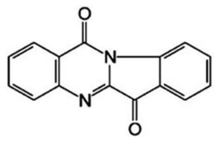
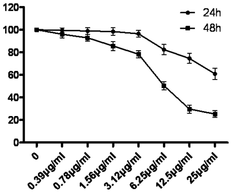
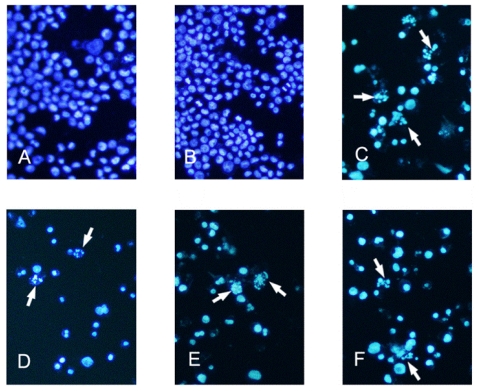
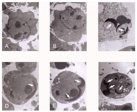
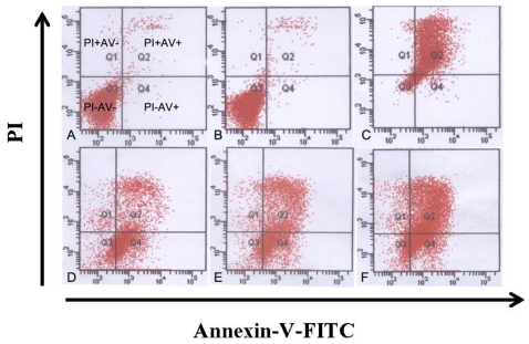
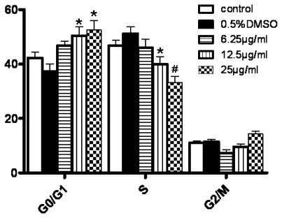
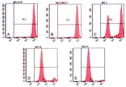
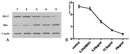
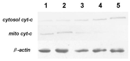
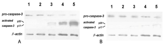
Tryptanthrin, a kind of indole quinazoline alkaloid, has been shown to exhibit anti-microbial, anti-inflammation and anti-tumor effects both in vivo and in vitro. However, its biological activity on human chronic myeloid leukemia cell line K562 is not fully understood. In the present study, we investigated the proliferation-attenuating and apoptosis-inducing effects of tryptanthrin on leukemia K562 cells in vitro and explored the underlying mechanisms. The results showed that tryptanthrin could significantly inhibit K562 cells proliferation in a time- and dose-dependent manner as evidenced by MTT assay and flow cytometry analysis. We also observed pyknosis, chromatin margination and the formation of apoptotic bodies in the presence of tryptanthrin under the electron microscope. Nuclei fragmentation and condensation by Hoechst 33258 staining were detected as well. The amount of apoptotic cells significantly increased whereas the mitochondrial membrane potential decreased dramatically after tryptanthrin exposure. K562 cells in the tryptanthrin treated group exhibited an increase in cytosol cyt-c, Bax and activated caspase-3 expression while a decrease in Bcl-2, mito cyt-c and pro-caspase-3 contents. However, the changes of pro-caspase-3 and activated caspase-3 could be abolished by a pan-caspase inhibitor ZVAD-FMK. These results suggest that tryptanthrin has proliferation-attenuating and apoptosis-inducing effects on K562 cells. The underlying mechanism is probably attributed to the reduction in mitochondria membrane potential, the release of mito cyt-c and pro-caspase-3 activation.

Keywords: K562 cells; apoptosis; chronic myeloid leukemia; proliferation; tryptanthrin.


Figures

Figure 1
Figure 1
Chemical structure of tryptanthrin.
Figure 2
Figure 2
Effect of tryptanthrin on the survival rate of K562 cells. Cells were treated with various concentrations of tryptanthrin for 24 h and 48 h. The cell survival rate was determined by MTT assay. Each data point is the mean of three replicates; bars represent the standard deviation.
Figure 3
Figure 3
Fluorescent staining of nuclei in tryptanthrin-treated K562 cells by Hoechst 33258. Cells were incubated in the medium without tryptanthrin (A) or with 0.5% DMSO (B), CTX (C) and with 6.25, 12.5, 25 μg/mL tryptanthrin (D, E and F) for 48 h, respectively. Fragmented or condensed nuclei could be observed at 200× magnification in the tryptanthrin-treated group as indicated by the arrows.
Figure 4
Figure 4
Transmission electron micrographs of K562 cells treated with different concentrations of tryptanthrin. No abnormal changes were observed in the control (A) and 0.5% DMSO (B) groups. Pyknosis, chromatin margination and the formation of apoptotic bodies (white arrows) were clearly observed in the presence of CTX (C) and tryptanthrin at the concentrations of 6.25 (D), 12.5 (E) and 25 (F) μg/mL, respectively.
Figure 5
Figure 5
Tryptanthrin caused strong K562 cells apoptosis. Representative FCM analysis scattergrams of Annexin V/PI stained control, 0.5% DMSO and treatment with CTX and 6.25, 12.5 and 25 μg/mL tryptanthrin for 48 h cells showed four different cell populations marked as: live cell population (PI−AV−), early apoptosis (PI−AV+), late apoptosis (PI+AV+) and dead cells (PI+AV−). Cells treated with no tryptanthrin (A), 0.5% DMSO (B), CTX (C) and 6.25, 12.5 and 25μg/mL tryptanthrin, respectively (D, E and F).
Figure 6
Figure 6
Cell cycle distribution analysis after tryptanthrin exposure. The proportions of G0/G1, S and G2/M phase cells were measured by Modfit 3.0 program. * P < 0.05, # P < 0.01.
Figure 7
Figure 7
Induction of K562 cell mitochondrial membrane collapse in the presence of tryptanthrin. Mitochondrial membrane potential was assessed by FCM after Rhodamine 123 staining. Rhodamine 123 enriched mainly in mitochondria showing red fluorescence reflecting changes of mitochondrial membrane potential. Cells were treated with no tryptanthrin (A), 0.5% DMSO (B) and 6.25, 12.5 and 25μg/mL tryptanthrin, respectively (C, D and E).
Figure 8
Figure 8
Western blot assay of Bcl-2, Bax and β-actin protein expression in K562 cells. (A) expression of Bcl-2 and Bax after tryptanthrin exposure. β-actin was used for normalization and verification of protein loading; (B) the Bcl-2/Bax ratio in the control, 0.5% DMSO and each tryptanthrin-treated groups. Lane 1: control; lane 2: 0.5% DMSO; lane 3: 6.25 μg/mL tryptanthrin; lane 4: 12.5 μg/mL tryptanthrin; lane 5: 25 μg/mL tryptanthrin.
Figure 9
Figure 9
Western blot assay of cytosol cyt-c and mito cyt-c in K562 cells. β-actin was used as an internal control. The expressions of cytosol cyt-c were very low in the control and 0.5% DMSO group. Tryptanthrin administration could lead to mito cyt-c leakage into the cytoplasm and increase cytosol contents in a dose dependent manner. Lane 1: control; lane 2: 0.5%DMSO; lane 3: 6.25 μg/mL tryptanthrin; lane 4: 12.5 μg/mL tryptanthrin; lane 5: 25 μg/mL tryptanthrin.
Figure 10
Figure 10
Western blot assay of pro-caspase-3 and activated caspase-3 in K562 cells. β-actin was used as an internal control. Caspase-3 exists as its inactive form pro-caspase-3 and there are no detectable active caspase-3 subunits in K562 cells. Administration of tryptanthrin induces mito cyt-c leakage and activates pro-caspase-3. The expression of active caspase-3 subunits p20 and p17 were remarkably elevated while pro-caspase-3 contents decreased after tryptanthrin exposure. However, addition of a pan-caspase inhibitor ZVAD-FMK significantly attenuated the pro-caspase-3 activation in response to tryptanthrin. The contents of p20 and p17 remarkably decreased in the presence of ZVAD-FMK. (A) Lane 1: control; lane 2: 0.5%DMSO; lane 3: 6.25 μg/mL tryptanthrin; lane 4: 12.5 μg/mL tryptanthrin; lane 5: 25 μg/mL tryptanthrin; (B) Lane 1: control; lane 2: ZVAD-FMK; lane 3: ZVAD-FMK + 6.25 μg/mL tryptanthrin; lane 4: ZVAD-FMK + 12.5 μg/mL tryptanthrin; lane 5: ZVAD-FMK + 25 μg/mL tryptanthrin.

Similar articles

Cited by

References

    1. Liu JJ, Zhang Y, Guang WB, Yang HZ, Lin DJ, Xiao RZ. Ponicidin inhibits monocytic leukemia cell growth by induction of apoptosis. Int. J. Mol. Sci. 2008;9:2265–2277. - PMC - PubMed
    1. Shim MJ, Kim HJ, Yang SJ, Lee IS, Choi HI, Kim T. Arsenic trioxide induces apoptosis in chronic myelogenous leukemia K562 cells: Possible involvement of p38 MAP kinase. J. Biochem. Mol. Biol. 2002;35:377–383. - PubMed
    1. Zhang Y, Dawson MI, Mohammad R, Rishi AK, Farhana L, Feng KC, Leid M, Peterson V, Zhang XK, Edelstein M, et al. Induction of apoptosis of human B-CLL and ALL cells by a novel retinoid and its nonretinoidal analog. Blood. 2002;100:2917–2925. - PubMed
    1. Chowdhury I, Tharakan B, Bhat GK. Current concepts in apoptosis: The physiological suicide program revisited. Cell. Mol. Biol. Lett. 2006;11:506–525. - PMC - PubMed
    1. Darzynkiewicz Z, Juan G, Li X, Gorczyca W, Murakami T, Traganos F. Cytometry in cell necrobiology: Analysis of apoptosis and accidental cell death (necrosis) Cytometry. 1997;27:1–20. - PubMed

Publication types

MeSH terms