. 2011 Apr 18:2:78.
doi: 10.3389/fmicb.2011.00078. eCollection 2011.

Intra-Vacuolar Proliferation of F. Novicida within H. Vermiformis

Affiliations

Intra-Vacuolar Proliferation of F. Novicida within H. Vermiformis

Marina Santic et al. Front Microbiol. .

Abstract

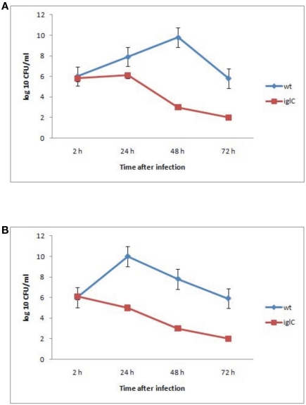
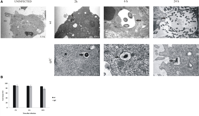
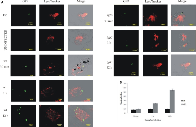
Francisella tularensis is a gram negative facultative intracellular bacterium that causes the zoonotic disease tularemia. Free-living amebae, such as Acanthamoeba and Hartmannella, are environmental hosts of several intracellular pathogens. Epidemiology of F. tularensis in various parts of the world is associated with water-borne transmission, which includes mosquitoes and amebae as the potential host reservoirs of the bacteria in water resources. In vitro studies showed intracellular replication of F. tularensis within A. castellanii cells. Whether ameba is a biological reservoir for Francisella in the environment is not known. We used Hartmannella vermiformis as an amebal model system to study the intracellular life of F. novicida. For the first time we show that F. novicida survives and replicates within H. vermiformis. The iglC mutant strain of F. novicida is defective for survival and replication not only within A. castellanii but also in H. vermiformis cells. In contrast to mammalian cells, where bacteria replicate in the cytosol, F. novicida resides and replicates within membrane-bound vacuoles within the trophozoites of H. vermiformis. In contrast to the transient residence of F. novicida within acidic vacuoles prior to escaping to the cytosol of mammalian cells, F. novicida does not reside transiently or permanently in an acidic compartment within H. vermiformis when examined 30 min after initiation of the infection. We conclude that F. tularensis does not replicate within acidified vacuoles and does not escape into the cytosol of H. vermiformis. The Francisella pathogenicity island locus iglC is essential for intra-vacuolar proliferation of F. novicida within H. vermiformis. Our data show a distinct intracellular lifestyle for F. novicida within H. vermiformis compared to mammalian cells.

Keywords: Francisella novicida; Hartmannella vermiformis; LysoTracker; iglC; vacuolar replication.


Figures

Figure 1
Figure 1
Iintracellular growth of F. novicida and its isogenic mutant iglC in A. castellanii (A) and in H. vermiformis (B). The cells were infected for 15 min, followed by gentamicin treatment, and determination of the number of intracellular bacteria at the indicated time points. The error bars represent standard deviations of triplicate samples and results shown are representative of three independent experiments.
Figure 2
Figure 2
Francisella novicida replicates in vacuoles of H. vermiformis. (A) Representative electron micrographs of H. vermiformis infected with wt F. tularensis subsp. novicida or the iglC mutant at 2, 6, and 24 h after infection. Thin black arrows show intact vacuolar membranes and white arrows show bacteria. (B) Quantitative analysis of the integrity of vacuolar membranes containing wt F. novicida or iglC in H. vermiformis cells. The percentage of disrupted vacuoles harboring bacteria at different time points.
Figure 3
Figure 3
Francisella novicida does not reside in acidic compartments within H. vermiformis cells. (A) Representative confocal microscopy images of colocalization of FCVs with the LysoTracker dye by the GFP-expressing wt F. novicida and iglC at 30 min, 1, and 12 h post-infection is shown. Uninfected cells were used as a negative control, while formalin killed bacteria served as a positive control. The images are representatives of 100 infected cells examined from three different cover slips. The results shown are representative of three independent experiments. (B) Quantification of colocalization of the LysoTracker DND-99 dye with the FCVs of the bacterium at 30 min, 1, and 12 h post-infection is shown. The results shown are representative of three independent experiments, and error bars represent standard deviations of triplicate samples.

References

    1. Abd H., Johansson T., Golovliov I., Sandstrom G., Forsman M. (2003). Survival and growth of Francisella tularensis in Acanthamoeba castellanii. Appl. Environ. Microbiol. 69, 600–606 - PMC - PubMed
    1. Abu Kwaik Y. (1996). The phagosome containing Legionella pneumophila within the protozoan Hartmannella vermiformis is surrounded by the rough endoplasmic reticulum. Appl. Environ. Microbiol. 62, 2022–2028 - PMC - PubMed
    1. Abu Kwaik Y., Gao L.-Y., Stone B. J., Venkataraman C., Harb O. S. (1998). Invasion of protozoa by Legionella pneumophila and its role in bacterial ecology and pathogenesis. Appl. Environ. Microbiol. 64, 3127–3133 - PMC - PubMed
    1. Akimana C., Abu Kwaik Y. (2011). Francisella–arthropod vector interaction and its role in patho-adaptation to infect mammals. Front. Microbio. 2:34.10.3389/fmicb.2011.00034 - DOI - PMC - PubMed
    1. Amann R., Springer N., Schonhuber W., Ludwig W., Schmid E. N., Muller K.-D., Michel R. (1997). Obligate intracellular bacterial parasites of Acanthamoebae related to Chlamydia spp. Appl. Environ. Microbiol. 63, 115–121 - PMC - PubMed

LinkOut - more resources