

Subversion of mucosal barrier polarity by pseudomonas aeruginosa
- PMID: 21747810
- PMCID: PMC3129012
- DOI: 10.3389/fmicb.2011.00114
Subversion of mucosal barrier polarity by pseudomonas aeruginosa
Abstract
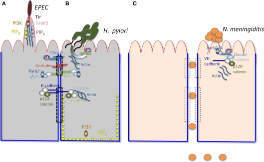
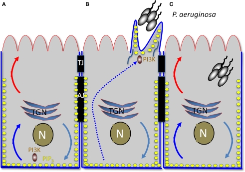
The lumenal surfaces of human body are lined by a monolayer of epithelia that together with mucus secreting cells and specialized immune cells form the mucosal barrier. This barrier is one of the most fundamental components of the innate immune system, protecting organisms from the vast environmental microbiota. The mucosal epithelium is comprised of polarized epithelial cells with distinct apical and basolateral surfaces that are defined by unique set of protein and lipid composition and are separated by tight junctions. The apical surface serves as a barrier to the outside world and is specialized for the exchange of materials with the lumen. The basolateral surface is adapted for interaction with other cells and for exchange with the bloodstream. A wide network of proteins and lipids regulates the formation and maintenance of the epithelium polarity. Many human pathogens have evolved virulence mechanisms that target this network and interfere with epithelial polarity to enhance binding to the apical surface, enter into cells, and/or cross the mucosal barrier. This review highlights recent advances in our understanding of how Pseudomonas aeruginosa, an important opportunistic human pathogen that preferentially infects damaged epithelial tissues, exploits the epithelial cell polarization machinery to enhance infection.
Keywords: Pseudomonas aeruginosa; adherens junctions; cell polarity; epithelial barrier; host–pathogen interactions; microbial pathogenesis; tight junctions.
Figures
Similar articles
-
Targeting the Mucosal Barrier: How Pathogens Modulate the Cellular Polarity Network.Cold Spring Harb Perspect Biol. 2017 Jun 1;9(6):a027953. doi: 10.1101/cshperspect.a027953. Cold Spring Harb Perspect Biol. 2017. PMID: 28193722 Free PMC article. Review.
-
Super-resolution imaging uncovers the nanoscopic segregation of polarity proteins in epithelia.Elife. 2022 Nov 7;11:e62087. doi: 10.7554/eLife.62087. Elife. 2022. PMID: 36341714 Free PMC article.
-
Epithelial cell polarity alters Rho-GTPase responses to Pseudomonas aeruginosa.Mol Biol Cell. 2004 Feb;15(2):411-9. doi: 10.1091/mbc.e03-08-0559. Epub 2003 Oct 31. Mol Biol Cell. 2004. PMID: 14595106 Free PMC article.
-
Pseudomonas aeruginosa-mediated damage requires distinct receptors at the apical and basolateral surfaces of the polarized epithelium.Infect Immun. 2010 Mar;78(3):939-53. doi: 10.1128/IAI.01215-09. Epub 2009 Dec 14. Infect Immun. 2010. PMID: 20008530 Free PMC article.
-
Modulation of epithelial cell polarity by bacterial pathogens.Ann N Y Acad Sci. 2017 Oct;1405(1):16-24. doi: 10.1111/nyas.13388. Epub 2017 Jun 19. Ann N Y Acad Sci. 2017. PMID: 28628193 Free PMC article. Review.
Cited by
-
The T6SSs of Pseudomonas aeruginosa Strain PAO1 and Their Effectors: Beyond Bacterial-Cell Targeting.Front Cell Infect Microbiol. 2016 Jun 9;6:61. doi: 10.3389/fcimb.2016.00061. eCollection 2016. Front Cell Infect Microbiol. 2016. PMID: 27376031 Free PMC article. Review.
-
Integrins as a bridge between bacteria and cells: key targets for therapeutic wound healing.Burns Trauma. 2024 Jul 16;12:tkae022. doi: 10.1093/burnst/tkae022. eCollection 2024. Burns Trauma. 2024. PMID: 39015251 Free PMC article. Review.
-
Modeling Host-Pathogen Interactions in the Context of the Microenvironment: Three-Dimensional Cell Culture Comes of Age.Infect Immun. 2018 Oct 25;86(11):e00282-18. doi: 10.1128/IAI.00282-18. Print 2018 Nov. Infect Immun. 2018. PMID: 30181350 Free PMC article. Review.
-
Pouring salt on a wound: Pseudomonas aeruginosa virulence factors alter Na+ and Cl- flux in the lung.J Bacteriol. 2013 Sep;195(18):4013-9. doi: 10.1128/JB.00339-13. Epub 2013 Jul 8. J Bacteriol. 2013. PMID: 23836869 Free PMC article. Review.
-
2,4-Di-Tert-Butylphenol Isolated From an Endophytic Fungus, Daldinia eschscholtzii, Reduces Virulence and Quorum Sensing in Pseudomonas aeruginosa.Front Microbiol. 2020 Jul 27;11:1668. doi: 10.3389/fmicb.2020.01668. eCollection 2020. Front Microbiol. 2020. PMID: 32849344 Free PMC article.
References
-
- Alaoui-El-Azher M., Jia J., Lian W., Jin S. (2006). ExoS of Pseudomonas aeruginosa induces apoptosis through a Fas receptor/caspase 8-independent pathway in HeLa cells. Cell. Microbiol. 8, 326–338 - PubMed
-
- Allesen-Holm M., Barken K. B., Yang L., Klausen M., Webb J. S., Kjelleberg S., Molin S., Givskov M., Tolker-Nielsen T. (2006). A characterization of DNA release in Pseudomonas aeruginosa cultures and biofilms. Mol. Microbiol. 59, 1114–1128 - PubMed
-
- Barrila J., Radtke A. L., Crabbe A., Sarker S. F., Herbst-Kralovetz M. M., Ott C. M., Nickerson C. A. (2010). Organotypic 3D cell culture models: using the rotating wall vessel to study host–pathogen interactions. Nat. Rev. Microbiol. 8, 791–801 - PubMed
Grants and funding
LinkOut - more resources
Full Text Sources