. 2011 May 23:2:115.
doi: 10.3389/fmicb.2011.00115. eCollection 2011.

Rhesus M1.3S Cells Suitable for Biological Evaluation of Macaque-Tropic HIV/SIV Clones

Affiliations

Rhesus M1.3S Cells Suitable for Biological Evaluation of Macaque-Tropic HIV/SIV Clones

Naoya Doi et al. Front Microbiol. .
No abstract available


Figures

Figure 1
Figure 1
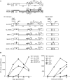
Virological characteristics of HIV-1/SIVmac clones. (A) Proviral genome structure of various viruses used for infection of M1.3S. Genomes of various HIV-1mt clones generated in our laboratory are schematically illustrated. Parental clones HIV-1 NL4-3 (Adachi et al., 1986) and SIVmac MA239N (Doi et al., 2010) are shown at the top. Accessory gene-inactivated mutants of SIVmac MA239N (circled) were constructed by substituting an internal stop codon in the nef gene of MA239 mutants (Shibata et al., 1991) with a Glu codon as reported for construction of MA239N (Doi et al., 2010). White and gray areas indicate the genomic regions of NL4-3 and MA239N, respectively. An arrow and arrowheads indicate the site of the four mutations in gag, pol, and env genes, respectively. The dotted region contains additional single-nucleotide mutations relative to the other clones. Enlarged Gag-CA region is shown on the right. For details, see the text. (B) Replication kinetics of the viruses in M1.3S cells. Cell-free viruses were prepared from 293T cells transfected with proviral clones indicated, and inoculated into 1 × 106 cells of M1.3S. Viral replication was monitored at intervals by reverse transcriptase (RT) activity in the culture supernatants. The experiments were done as described previously (Kamada et al., ; Doi et al., 2010). For infection, 6.3 × 104 and 2.4 × 106 RT units of MA239N and HIV-1mt clones (left), respectively, and 5.0 × 104 RT units of MA239N clones (right) were used. The representative data in two independent experiments are shown. Replication of NL-DT5RS, MN4–8, and MN4–8S was not detected as observed for NL-DT5R.

References

    1. Adachi A., Gendelman H. E., Koenig S., Folks T., Willey R., Rabson A., Martin M. A. (1986). Production of acquired immunodeficiency syndrome-associated retrovirus in human and nonhuman cells transfected with an infectious molecular clone. J. Virol. 59, 284–291 - PMC - PubMed
    1. Akari H., Mori K., Terao K., Otani I., Fukasawa M., Mukai R., Yoshikawa Y. (1996). In vitro immortalization of Old World monkey T lymphocytes with Herpesvirus saimiri: its susceptibility to infection with simian immunodeficiency viruses. Virology 218, 382–38810.1006/viro.1996.0207 - DOI - PubMed
    1. Ambrose Z., KewalRamani V. N., Bieniasz P. D., Hatziioannou T. (2007). HIV/AIDS: in search of an animal model. Trends Biotechnol. 25, 333–33710.1016/j.tibtech.2007.05.004 - DOI - PubMed
    1. Doi N., Fujiwara T., Adachi A., Nomaguchi M. (2010). Growth ability in various macaque cell lines of HIV-1 with simian cell-tropism. J. Med. Invest. 57, 284–292 - PubMed
    1. Fujita M., Yoshida A., Sakurai A., Tatsuki J., Ueno F., Akari H., Adachi A. (2003). Susceptibility of HVS-immortalized lymphocytic HSC-F cells to various strains and mutants of HIV/SIV. Int. J. Mol. Med. 11, 641–644 - PubMed

LinkOut - more resources