Cutaneous squamous cell carcinoma (SCC) and the DNA damage response: pATM expression patterns in pre-malignant and malignant keratinocyte skin lesions
- PMID: 21747934
- PMCID: PMC3128585
- DOI: 10.1371/journal.pone.0021271
Cutaneous squamous cell carcinoma (SCC) and the DNA damage response: pATM expression patterns in pre-malignant and malignant keratinocyte skin lesions
Abstract
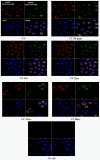
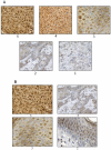
Recent evidence suggests that an initial barrier to the emergence of tumours is a DNA damage response that evokes a counter-response which arrests the growth of, or eliminates, damaged cells. Early precursor lesions express markers of an activated DNA damage response in several types of tumour, with a diminishing response in more advanced cancers. An important marker of DNA damage is ATM which becomes phosphorylated (pATM) upon activation. We have investigated pATM expression patterns in cultured keratinocytes, skin explants and a spectrum of pre-malignant to malignant keratinocyte skin lesions by immunohistochemistry. We found that pATM was mainly localised to the Golgi apparatus, which contrasts with its nuclear localisation in other tissues. Upon UV irradiation there is transient formation of pATM in nuclear foci, consistent with recruitment to the sites of DNA damage. By immunohistochemistry we show pATM expression in precancerous keratinocyte lesions is greater and predominantly nuclear when compared to the invasive lesions where pATM is weaker and predominantly cytoplasmic. Our results are consistent with the hypothesis that the DNA damage response acts as a barrier to cutaneous tumour formation, but also suggests that ATM expression in skin is different compared to other tissues. This may be a consequence of the constant exposure of skin to UVR, and has implications for skin carcinogenesis.
Conflict of interest statement
Figures


Comment in
-
Editorial Note: Cutaneous Squamous Cell Carcinoma (SCC) and the DNA Damage Response: pATM Expression Patterns in Pre-Malignant and Malignant Keratinocyte Skin Lesions.PLoS One. 2024 May 2;19(5):e0303120. doi: 10.1371/journal.pone.0303120. eCollection 2024. PLoS One. 2024. PMID: 38696417 Free PMC article. No abstract available.
References
-
- Norbury CJ, Hickson ID. Cellular responses to DNA damage. Annu Rev Pharmacol Toxicol. 2001;41:367–401. - PubMed
-
- Bakkenist CJ, Kastan MB. DNA damage activates ATM through intermolecular autophosphorylation and dimer dissociation. Nature. 2003;421:499–506. - PubMed
-
- Gorgoulis VG, Vassiliou LV, Karakaidos P, Zacharatos P, Kotsinas A, et al. Activation of the DNA damage checkpoint and genomic instability in human precancerous lesions. Nature. 2005;434:907–913. - PubMed
-
- Bartkova J, Horejsi Z, Koed K, Kramer A, Tort F, et al. DNA damage response as a candidate anti-cancer barrier in early human tumorigenesis. Nature. 2005;434:864–870. - PubMed
-
- Bartek J, Bartkova J, Lukas J. DNA damage signalling guards against activated oncogenes and tumour progression. Oncogene. 2007;26:7773–7779. - PubMed
Publication types
MeSH terms
Substances
Grants and funding
LinkOut - more resources
Full Text Sources
Medical
Molecular Biology Databases
Research Materials
Miscellaneous

