

Grey-blue regression in melanoma in situ-evaluation on 111 cases
- PMID: 21748024
- PMCID: PMC3118612
- DOI: 10.1155/2011/180980
Grey-blue regression in melanoma in situ-evaluation on 111 cases
Abstract
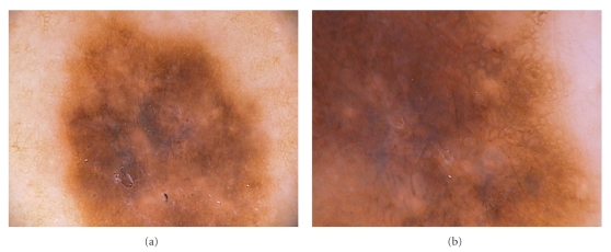
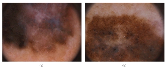
As fibrosis and melanosis are often seen in malignant melanoma, the presence of dermoscopic signs of regression may represent a clue for the diagnosis of malignancy. Our aim was to assess the frequency and extent of 11 dermoscopic features of regression evaluating dermoscopic images of 111 melanomas in situ (MIS). Regression structures (grey-blue areas, white areas, peppering, and/or blue-whitish veil) were present in 80.1% of the lesions. Approximately 80% of the lesions showed regression of dermoscopic structures and light brown areas. Most lesions showed the presence of grey-blue areas (74.7%), whereas peppering was observable in 30.6% of MIS. Areas of fibrosis were mainly observable as structureless areas with a pinkish hue (50.4%). Based on our data, the reticular pattern of blue regression and light brown areas can be considered a significant discriminator and a reliable predictor of MIS.
Figures
References
-
- Pizzichetta MA, Talamini R, Stanganelli I, et al. Amelanotic/hypomelanotic melanoma: clinical and dermoscopic features. British Journal of Dermatology. 2004;150(6):1117–1124. - PubMed
-
- Argenziano G, Fabbrocini G, Carli P, De Giorgi V, Sammarco E, Delfino M. Epiluminescence microscopy for the diagnosis of doubtful melanocytic skin lesions: comparison of the ABCD rule of dermatoscopy and a new 7-point checklist based on pattern analysis. Archives of Dermatology. 1998;134(12):1563–1570. - PubMed
-
- Zalaudek I, Argenziano G, Ferrara G, et al. Clinically equivocal melanocytic skin lesions with features of regression: a dermoscopic-pathological study. British Journal of Dermatology. 2004;150(1):64–71. - PubMed
-
- Argenziano G, Soyer HP, Chimenti S, et al. Dermoscopy of pigmented skin lesions: results of a consensus meeting via the internet. Journal of the American Academy of Dermatology. 2003;48(5):679–693. - PubMed
-
- Argenziano G, Soyer HP, De Giorgi V, et al. Interactive Atlas of Dermoscopy. Milan, Italy: Edra Medical Publishing and New Media; 2000.
LinkOut - more resources
Full Text Sources
Research Materials
