Comparative Study
. 2012 Jan;64(1):49-58.
doi: 10.1007/s00251-011-0558-0. Epub 2011 Jul 7.

Comparative genomic analysis of the proteasome β5t subunit gene: implications for the origin and evolution of thymoproteasomes

Affiliations
Comparative Study

Comparative genomic analysis of the proteasome β5t subunit gene: implications for the origin and evolution of thymoproteasomes

Yoichi Sutoh et al. Immunogenetics. 2012 Jan.

Abstract

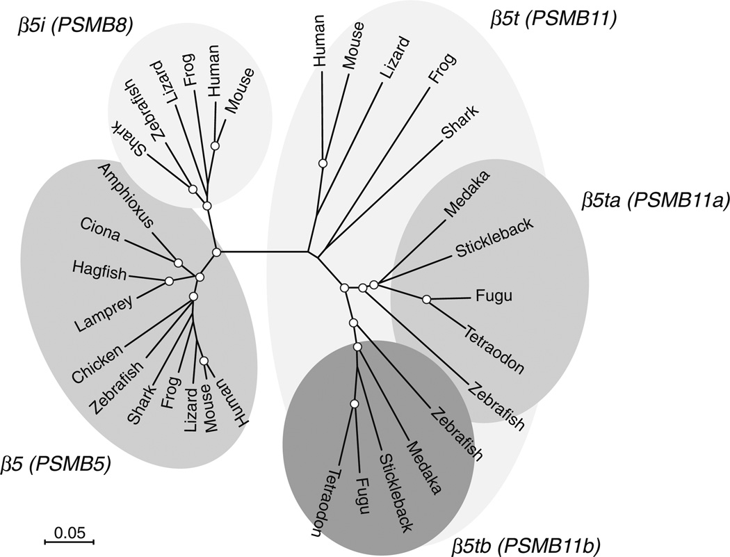
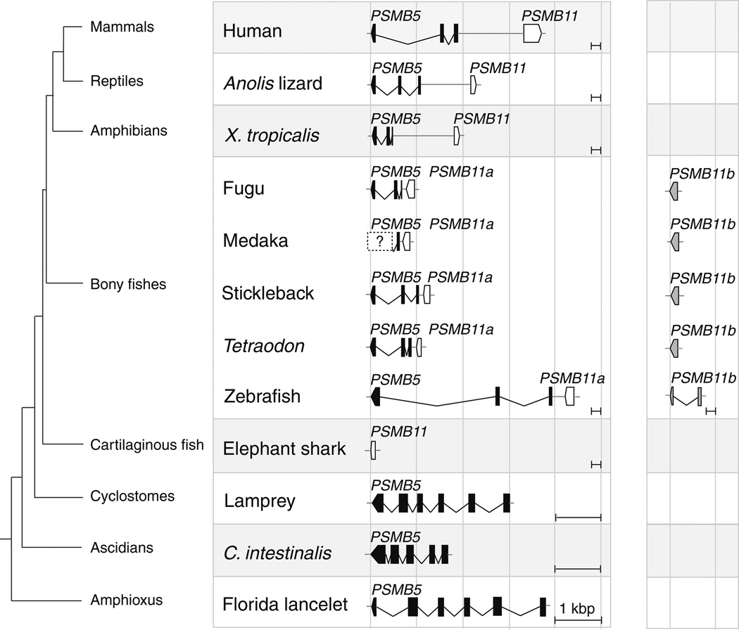
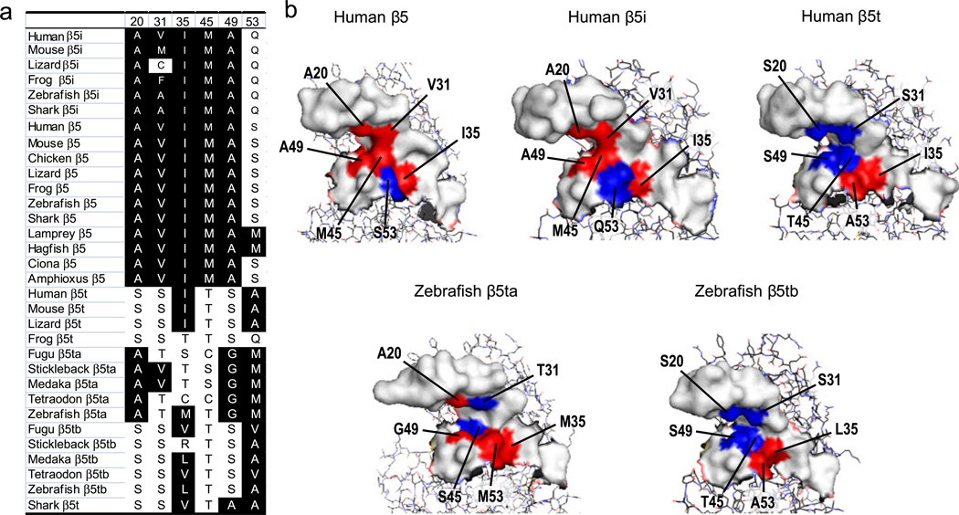
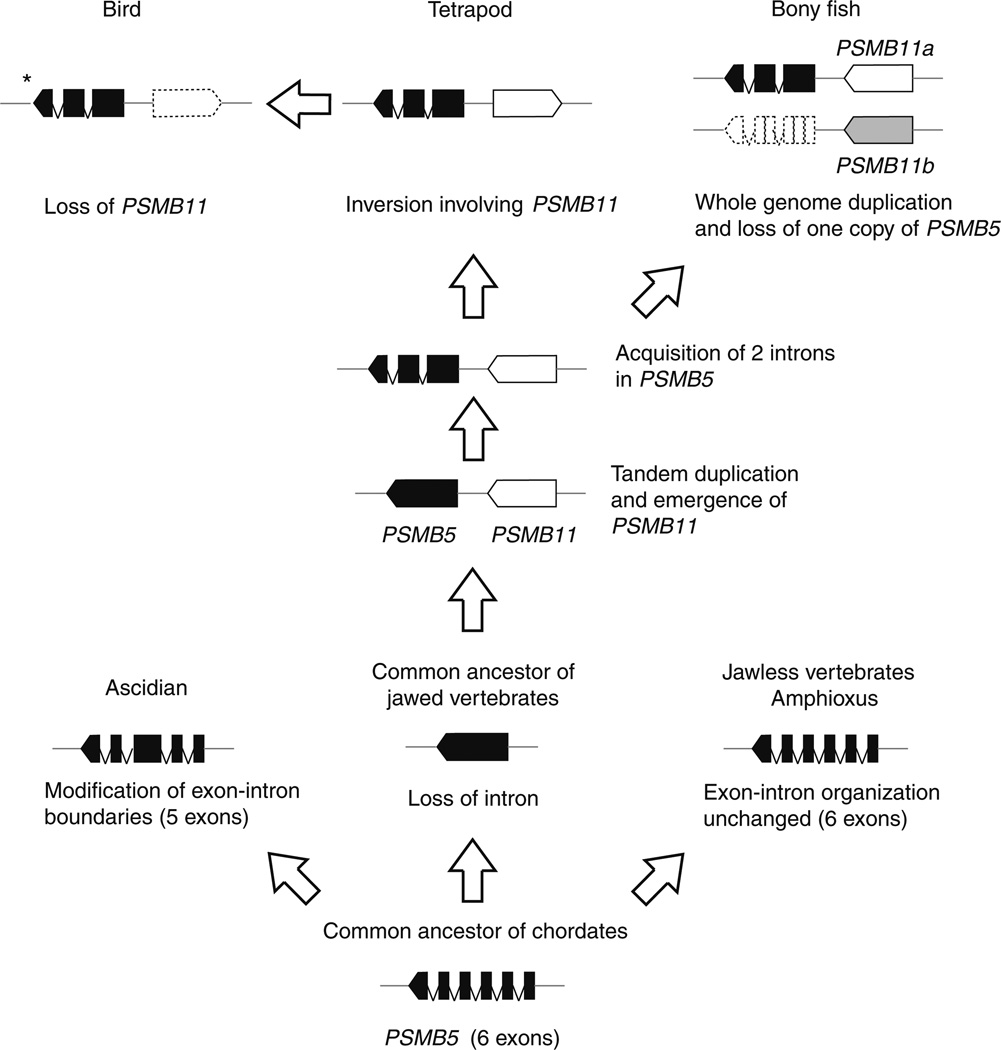
The thymoproteasome is a recently discovered, specialized form of 20S proteasomes expressed exclusively in the thymic cortex. Although the precise molecular mechanism by which the thymoproteasome exerts its function remains to be elucidated, accumulating evidence indicates that it plays a crucial role in positive selection of T cells. In the present study, we analyzed the evolution of the β5t subunit, a β-type catalytic subunit uniquely present in thymoproteasomes. The gene coding for the β5t subunit, designated PSMB11, was identified in the cartilaginous fish, the most divergent group of jawed vertebrates compared to the other jawed vertebrates, but not in jawless vertebrates or invertebrates. Interestingly, teleost fish have two copies of apparently functional PSMB11 genes, designated PSMB11a and PSMB11b, that encode β5t subunits with distinct amino acids in the S1 pocket. BLAST searches of genome databases suggest that birds such as chickens, turkey, and zebra finch lost the PSMB11 gene, and have neither thymoproteasomes nor immunoproteasomes. In mammals, reptiles, amphibians, and teleost fishes, the PSMB11 gene (the PSMB11a gene in teleost fish) is located next to the PSMB5 gene coding for the β5 subunit of the standard 20S proteasome, indicating that the PSMB11 gene arose by tandem duplication from the evolutionarily more ancient PSMB5 gene. The general absence of introns in PSMB11 and an unusual exon-intron structure of jawed vertebrate PSMB5 suggest that PSMB5 lost introns and duplicated in tandem in a common ancestor of jawed vertebrates, with PSMB5 subsequently gaining two introns and PSMB11 remaining intronless.


Figures

Fig. 1
Fig. 1
Phylogenetic tree of the β5 family of proteasome subunits. Nodes supported by bootstrap values over 90% are indicated by open circles. DDBJ/EMBL/NCBI accession numbers or Ensembl gene identification numbers are as follows: human β5i, P28062; mouse β5i, NP_034854.2; Anolis lizard β5i, ENSACAT00000006461; X. tropicalis β5i, NP_001103512.1; zebrafish β5i, AAB87679.1; nurse shark β5i, BAA10934.1; human β5, NP_002788.1; mouse β5, BAA24916.1; chicken β5, BAA19471.1; Anolis lizard β5, FG667020.1; X. tropicalis β5, XP_002941560.1; zebrafish β5, AAI53590.1; nurse shark β5, BAA10935.1; lamprey β5, BAA10932.1; hagfish β5, BAA10931.1; C. intestinalis β5, XP_002132107.1; Florida lancelet β5, AAM18885.1; human β5t, BAF63540.1; mouse β5t, BAF63539.1; Anolis lizard β5t, ENSA CAT00000001900; X. tropicalis β5t, XP_002941563.1; zebrafish β5ta, AB624351; zebrafish β5tb, AB624352; fugu β5ta, ENSTRUT00000004609; fugu β5tb, ENSTRUT00000034202; stickleback β5tb, ENSGACT00000002260; Tetraodon β5ta, ENST-NIT00000001930; Tetraodon β5tb, ENSTNIT00000009972; medaka β5tb, ENSORLT00000020386; and elephant shark β5t, AAVX01606762.1. Medaka and stickleback β5ta genes were not annotated in the databases
Fig. 2
Fig. 2
Genomic organization of PSMB11 and PSMB5 genes. Teleost fish have two PSMB11 genes located on different chromosomes or contigs. Only partial sequence data are available for the medaka PSMB5 gene. No avian PSMB11 sequence was identified in the NCBI/Ensembl databases. The Ensembl database contains only a partial genomic sequence of chicken PSMB5. Hence, its structure is not shown. Note that lamprey, Ciona, and Florida lancelet PSMB5 genes are drawn in scales different from those used for the remaining genes
Fig. 3
Fig. 3
The genomic regions containing PSMB11a and PSMB11b share paralogs of at least three other gene families. The Tetraodon genome contains two sets of closely related CDH24, LRRC16B, and TGM1 genes. One set, designated with suffix a, is located immediately downstream from the PSMB11a/PSMB5 gene cluster on choromosome 15. Another set of CDH24, LRRC16B, and TGM1 genes, designated with suffix b, is distributed between 11.20 and 12.09 Mbp on chromosome 1. PSMB11b is located at ~11.29 Mbp on choromosome 1. The information is based on the TETRAODON 8.0 assembly of the Ensembl database. Numbers starting with ENSTNIG are Ensembl gene identification numbers
Fig. 4
Fig. 4
Amino acid sequences forming the S1 pockets of β5i/β5/β5t subunits (a) and the predicted three-dimensional structures of the S1 pockets (b). In (a), hydrophobic and hybrophilic residues are boxed in black and white, respectively. In (b), hydrophobic and hybrophilic residues are indicated in red and blue, respectively. Amino acids are shown in a standard single-letter code
Fig. 5
Fig. 5
Evolution of β5 and β5t subunit genes. Exons and introns are indicated by filled boxes and lines, respectively. *Exon–intron organization of chicken PSMB5 is not known

Similar articles

Cited by

References

    1. Abdulla S, Beck S, Belich M, Jackson A, Nakamura T, Trowsdale J. Divergent intron arrangement in the MB1/LMP7 proteasome gene pair. Immunogenetics. 1996;44:254–258. - PubMed
    1. Arnold K, Bordoli L, Kopp J, Schwede T. The SWISS-MODEL workspace: a web-based environment for protein structure homology modelling. Bioinformatics. 2006;22:195–201. - PubMed
    1. Bajoghli B, Guo P, Aghaallaei N, Hirano M, Strohmeier C, McCurley N, Bockman DE, Schorpp M, Cooper MD, Boehm T. A thymus candidate in lampreys. Nature. 2011;470:90–94. - PubMed
    1. Balakrishnan CN, Ekblom R, Volker M, Westerdahl H, Godinez R, Kotkiewicz H, Burt DW, Graves T, Griffin DK, Warren WC, Edwards SV. Gene duplication and fragmentation in the zebra finch major histocompatibility complex. BMC Biol. 2010;8:29. - PMC - PubMed
    1. Belich MP, Trowsdale J. Proteasome and class I antigen processing and presentation. Mol Biol Rep. 1995;21:53–56. - PubMed

Publication types

MeSH terms

Associated data