. 2011 Oct;112(10):2759-72.
doi: 10.1002/jcb.23259.

Substance P signaling mediates BMP-dependent heterotopic ossification

Affiliations

Substance P signaling mediates BMP-dependent heterotopic ossification

Lixin Kan et al. J Cell Biochem. 2011 Oct.

Abstract

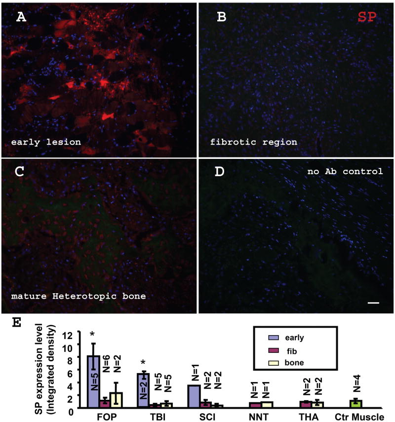
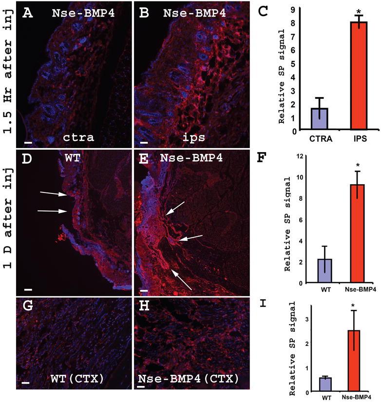
Heterotopic ossification (HO) is a disabling condition associated with neurologic injury, inflammation, and overactive bone morphogenetic protein (BMP) signaling. The inductive factors involved in lesion formation are unknown. We found that the expression of the neuro-inflammatory factor Substance P (SP) is dramatically increased in early lesional tissue in patients who have either fibrodysplasia ossificans progressiva (FOP) or acquired HO, and in three independent mouse models of HO. In Nse-BMP4, a mouse model of HO, robust HO forms in response to tissue injury; however, null mutations of the preprotachykinin (PPT) gene encoding SP prevent HO. Importantly, ablation of SP(+) sensory neurons, treatment with an antagonist of SP receptor NK1r, deletion of NK1r gene, or genetic down-regulation of NK1r-expressing mast cells also profoundly inhibit injury-induced HO. These observations establish a potent neuro-inflammatory induction and amplification circuit for BMP-dependent HO lesion formation, and identify novel molecular targets for prevention of HO.


Figures

Figure 1
Figure 1. SP is up-regulated in early FOP lesions
(A) SP up-regulation in an early FOP lesion. Note the widespread, but not universal, SP staining. Two patterns can be recognized: the strong punctuate and semi-diffuse staining. (B) No appreciable level of SP staining was observed in the fibroproliferative (pre-cartilage) stage. (C) shows the SP staining of the mature HO from a FOP patient. (D) Negative control (no 1st antibody). Bar=40 μm. (E) Summary of relative SP expression levels in FOP, acquired-HO patient, and normal controls. Relative SP expressions are graphed based on signal intensity of fluorescence images. fibrodysplasia ossificans progressiva (FOP), Spinal Cord Injury (SCI), Traumatic Brain Injury (TBI), Non-Neurologic Trauma (NNT) and Total Hip Arthroplasty (THA). Note significant SP up-regulation was observed only in early lesions. (* P<0.01 vs control muscles). Error bars represent s.d. early: early lesion, fib: fibrotic lesion, bone: mature bone.
Figure 2
Figure 2. SP staining is specific, and co-localized with a neuronal marker
(A&B) Peptide blocking experiment indicated that both punctuate and the semi-diffuse staining observed in early lesions are specific. (A) shows a typical image of SP staining in an early lesion from a FOP patient. (B) shows that both punctuated and the semi-diffused staining in a neighboring section are blocked by SP peptide. (C) shows a typical image of SP/NF-200 double staining in an early lesion from a traumatic brain injury (TBI) patient. Note that there is extensive co-localization of SP and NF-200 (white arrows in C). Also note that there are some NF-200 cells also express high level of SP (white arrow heads in C). (C′ and C″) shows the split channel of NF-200 and SP, respectively. (D) shows the typical image of SP stained in mature HO from a sample from a TBI patient. (E) shows the typical image of SP staining from a sample of normal control muscle. Bar=40 μm.
Figure 3
Figure 3. Injuries induced transgene dependent SP up-regulation in connective tissue and muscles
Both superficial muscles injury (A-F) and CTX induced deep muscles injury (G-I) induced SP up-regulation. (A&B) show typical images of SP immunostained cross sections of contralateral uninjured (A) and injured (B) limbs 1.5 hr after superficial muscles injury. Note the dramatic SP up-regulation and edema in the injured site (B). (C) quantified the relative SP signal in (A&B). (* P<0.01 vs control). Error bars represent s.d. (D&E) show typical images of SP immunostained cross sections of injured WT (D) and Nse-BMP4 (E) limbs one day after superficial muscles injury. Note the dramatic SP up-regulation and edema in Nse-BMP4 mice (E). (F) quantified the relative SP signal in (D&E). (* P<0.01 vs control). Error bars represent s.d. (G&H) show typical images of SP immunostained cross sections of injured WT (G) and Nse-BMP4 (H) limbs after CTX induced injury. (I) quantified the relative SP signal in (G&H). (* P<0.01 vs control). Error bars represent s.d. Bar=40 μm in A, B, G&H, Bar=200 μm in D&E.
Figure 4
Figure 4. Injury induced a unique immunohistochemical phenotype (mesh-like SP staining pattern) of lumbar DRGs of Nse-BMP4 mice
Sections of lumbar DRGs from age matched 6 month old WT (A&B) and Nse-BMP4 with HO (C&D) mice were immunostained for SP. A&C, untreated, and B&D, pretreated with colchcine (COL). Note that the unique mesh-like SP staining pattern was found only in Nse-BMP4 (C), and that the normal pattern was restored by colchcine treatment in Nse-BMP4 mice (D). (E) depicts the SP+ cell densities of different subpopulations of neurons in 6 month old Nse-BMP4 and age matched WT lumbar DRGs. Note that colchcine treatment increased the density of SP+ neurons in Nse-BMP4 DRGs without significantly changing the total number of P75+ neurons. (F&G) The mesh-like staining pattern is induced by superficial muscles injury (G, 1.5 hours after injury) in younger (one month old) Nse-BMP4 mice. (F) SP immunostained sections of lumbar DRGs from 1 month old naïve Nse-BMP4. Note that cytoplasmic staining of SP is still observed at this time point. White arrows point to the SP+ cell bodies in (G). NK1r antagonist (RP-67580) treatment had variable efficiency in restoring the normal staining pattern of SP in old Nse-BMP4 mice (H&I). SP stained sections of lumbar DRGs from 6 month old Nse-BMP4 treated with PBS (H), and 6 months old Nse-BMP4 pre-treated with NK1r antagonist for 2 weeks (I). Note that the normal staining pattern of SP in old Nse-BMP4 mice was only partially restored with NK1r antagonist treatment. White arrows point to the SP+ cells. Bar=40 μm.
Figure 5
Figure 5. Ablation of SP+ neurons inhibits injury induced SP up-regulation and inflammation
Small diameter SP+ sensory neurons were virtually absent in capsaicin pretreated Nse-BMP4 DRG (A) & (B), compared to vehicle treated ones (C)&(D). DRG depicted in (A&C) are from uninjured side, while (B & D) show the DRG from injured side. Note that injury did not lead to injury induced mesh-like pattern in capsaicin pretreated Nse-BMP4 mice (B), compared to vehicle treated one (D). Ablation of SP+ neurons inhibits injury-induced SP up-regulation in target tissues of Nse-BMP4 mice (E &F). Cross sections of hind limbs from the injury site are shown here. Vehicle (E) and capsaicin pretreated (F) mice were subjected to similar superficial muscles injury, hind limbs were harvested 1 hour after injury (shown) or 1 day after injury (not shown). Reduced SP up-regulation was observed in lesions from capsaicin pretreated Nse-BMP4 mice (F), compared to vehicle pretreated mice (E). The strong SP staining was found closely associated with muscle fibers. Infiltrating cells are indicated by dense DAPI+ regions. (G & H) H&E staining clearly indicated that ablation of SP+ neurons inhibited injury induced inflammatory response. (G) Image from injured site (1 hour after injury) of vehicle pretreated Nse-BMP4 mice demonstrated massive infiltration of inflammatory cells. In contrast, much less inflammatory cells infiltration was observed in injured site of capsaicin pretreated Nse-BMP4 mice (H). (I) quantified the relative SP levels observed in (E&F). * P<0.01 vs vehicle control. (J) Quantified the numbers of inflammatory cells observed in (G&H). * P<0.01 vs vehicle control.
Figure 6
Figure 6. Inhibition of SP signaling blocks injury induced inflammation and HO
(A) summarizes superficial muscles injury-induced edema and HO in different conditions. Nse-BMP4 mice without endogenous SP signaling (PPT-A−/−, Nk1r−/−, or ablation of SP+ neurons), or mast cells (c-kitw-sh/w-sh) did not develop injury induced HO efficiently. (B) Effect of SP receptor (NK1r) inhibition (RP-67580) in response to superficial muscles injury. Frequency (percent of mice) of HO formation over time (1-6 weeks) with RP-67580 or PBS treatment is shown. (C-G) RP-67580 inhibited HO in CTX induced muscle injury model. (C) shows the CTX induced HO (3D reconstruction) from a PBS treated Nse-BMP4 mouse. (D) shows that CTX induced HO was inhibited by RP-67580 treatment. (E) depicts the total HO volumes one month after muscles injury in RP-67580 and PBS treated groups. (F) shows gross images from the same groups. (G) depicts the total wet weights of affected hindlimbs in the same groups. * P<0.01 vs control. Note that the total HO volumes, total wet weights and gross images consistently indicated the inhibition effect of RP-67580. (H-K) RP-67580 specifically inhibited injury induced edema and inflammation. (H) shows gross images seven days after injury from RP-67580 and PBS treated limbs. Note that the edema in PBS treated limbs are much obvious than the limbs from RP-67580 group (reflected by circumferences). (I) depicts the total wet weights of affected hindlimbs from the same groups. * P<0.01 vs control. (J&K) show the typical images of H&E staining from RP-67580 and PBS treated group, respectively. Note that the inflammatory response is dramatically reduced in RP-67580 treated lesion (J), comparing to PBS (K) treated lesion.

References

    1. Salisbury E, Sonnet C, Heggeness M, Davis AR, Olmsted-Davis E. Heterotopic ossification has some nerve. Crit Rev Eukaryot Gene Expr. 2010;20:313–324. - PMC - PubMed
    1. Forsberg JA, Potter BK. Heterotopic ossification in wartime wounds. J Surg Orthop Adv. 2010;19:54–61. - PubMed
    1. Cullen N, Perera J. Heterotopic ossification: pharmacologic options. J Head Trauma Rehabil. 2009;24:69–71. - PubMed
    1. Shore EM, Xu M, Feldman GJ, Fenstermacher DA, Cho TJ, Choi IH, Connor JM, Delai P, Glaser DL, LeMerrer M, Morhart R, Rogers JG, Smith R, Triffitt JT, Urtizberea JA, Zasloff M, Brown MA, Kaplan FS. A recurrent mutation in the BMP type I receptor ACVR1 causes inherited and sporadic fibrodysplasia ossificans progressiva. Nat Genet. 2006;38:525–527. - PubMed
    1. Kaplan FS, Glaser DL, Shore EM, Pignolo RJ, Xu M, Zhang Y, Senitzer D, Forman SJ, Emerson SG. Hematopoietic stem-cell contribution to ectopic skeletogenesis. J Bone Joint Surg Am. 2007;89:347–357. - PubMed

Publication types

MeSH terms

LinkOut - more resources