. 2011 Jul 12;10(1):2.
doi: 10.1186/1476-5926-10-2.

Characterization of Kupffer cells in livers of developing mice

Affiliations

Characterization of Kupffer cells in livers of developing mice

Bryan G Lopez et al. Comp Hepatol. .

Abstract

Background: Kupffer cells are well known macrophages of the liver, however, the developmental characteristics of Kupffer cells in mice are not well understood. To clarify this matter, the characteristics of Kupffer macrophages in normal developing mouse liver were studied using light microscopy and immunocytochemistry.

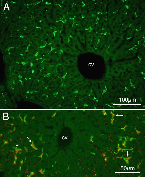
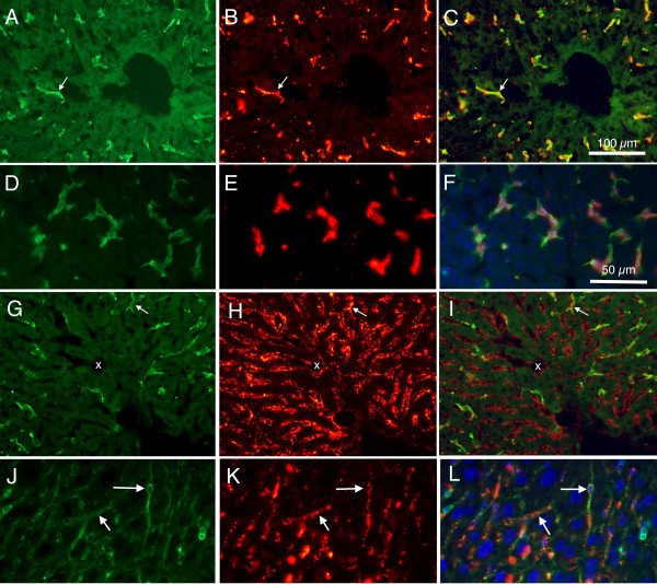
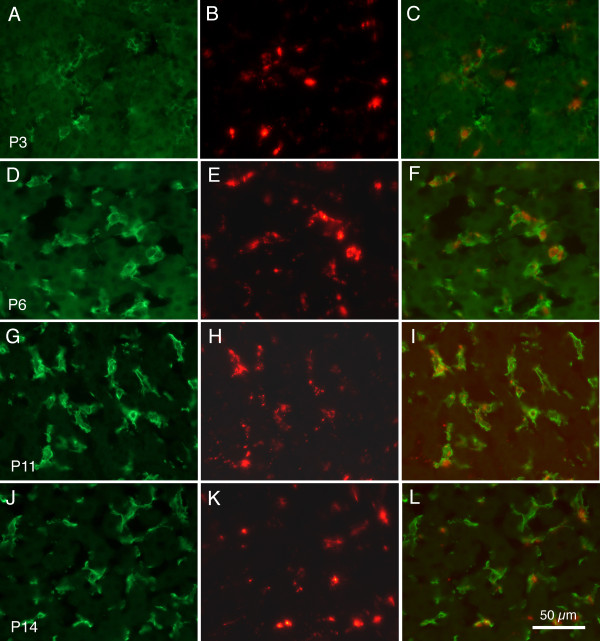
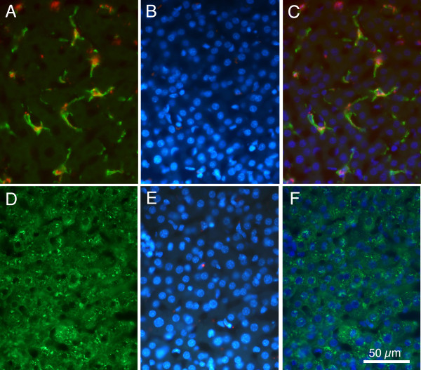
Methods: Sections of liver tissue from early postnatal mice were prepared using immunocytochemical techniques. The Kupffer cells were identified by their immunoreactivity to the F4/80 antibody, whereas endothelial cells were labelled with the CD-34 antibody. In addition, Kupffer cells and endothelial cells were labelled by systemically injected fluorescently labelled latex microspheres. Tissue slices were examined by fluorescence microscopy.

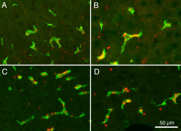
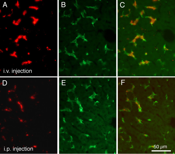
Results: Intravenous or intraperitonal injections of microspheres yielded similar patterns of liver cell labelling. The F4/80 positive Kupffer cells were labelled with both large (0.2 μm) and small (0.02 μm) diameter microspheres, while endothelial cells were labelled only with the smaller diameter microspheres. Microsphere labelling of Kupffer cells appeared stable for at least 6 weeks. Cells immunoreactive for F4/80 were identified as early as postnatal day 0, and these cells also displayed uptake of microspheres. Numbers of F4/80 Kupffer cells, relative to numbers of albumin positive hepatocytes, did not show a significant trend over the first 2 postnatal weeks.

Conclusions: Kupffer cells of the developing mouse liver appear quite similar to those of other mammalian species, confirming that the mouse presents a useful animal model for studies of liver macrophage developmental structure and function.


Figures

Figure 1
Figure 1
Fluorescence photomicrographs showing Kupffer cells from sections of P28 mouse liver. A: Alexa 488 (green) labelled F4/80 positive cells. Note branching of cells, and relative absence of positive cells close to the central venule (cv). Calibration bar = 100 μm. B: Merged image showing Alexa 488 (green) labelled F4/80 positive cells along with 0.2 μm red fluorescent microsphere positive cells. Arrows indicate examples of double labelled cells. Calibration bar = 50 μm.
Figure 2
Figure 2
Fluorescence photomicrographs from P30 and P15 mouse liver, showing difference in patterns of labeling between large (0.2 μm) and small (0.02) microspheres. A: Alexa 488 labelled F4/80 cells from P30 mouse. B: Same section as in 'A' but viewed using rhodamine optics to reveal large (0.2 μm) fluorescently labelled microspheres. C: Merged image of 'A' and 'B', showing co-localization of F4/80 and large microspheres. D: Higher magnification photomicrograph showing Alexa 488 labelled F4/80 cells from P15 mouse liver. E: Same section as in 'D', viewed using rhodamine optics to reveal large (0.2 μm) fluorescently labelled microspheres. F: Merged image of 'D' and 'E', and also with ultraviolet imaging of DAPI labelled cell nuclei, showing cells co-labelled with F4/80 and microspheres. Note that most microspheres appear associated with F4/80 positive cells. G: Alexa 488 labelled F4/80 positive cells from P30 mouse. H: Same section as in 'G', viewed using rhodamine optics to reveal small (0.02 μm) fluorescently labelled microspheres. I: Merged image of 'G' and 'H', showing a few cells co-labelled with F4/80 and microspheres. Note that most microspheres appear not to be associated with F4/80 positive cells. White arrows indicate double labelled cells; x indicates capillary with red microspheres but absence of F4/80 immunoreactivity. J: Higher magnification photomicrograph showing Alexa 488 labelled CD-34 cells from P15 mouse liver. K: Same section as in 'J', viewed using rhodamine optics to reveal small (0.02 μm) fluorescently labelled microspheres. L: Merged image of 'J' and 'K', and also with ultraviolet imaging of DAPI labelled cell nuclei, showing cells co-labelled with CD-34 and microspheres. Note that most microspheres appear associated with CD-34 positive cells; examples are indicated by white arrows. Calibration bar in 'C' = 100 μm for images A, B, C, G, H, and I. Calibration bar in 'F' = 50 μm for images D, E, F, J, K, and L.
Figure 3
Figure 3
Merged images of fluorescence photomicrographs from animals injected intravenously at P20 show Alexa 488 (green) labelled and large (0.2 μm) red fluorescent microsphere containing cells. A: 30 minutes following IV injection. B: 1 hr following injection. C: 1 week following injection. D: 2 weeks following injection. Calibration bar in 'D' = 50 μm for all images.
Figure 4
Figure 4
Fluorescence images allow comparison of results of IV and IP injections. Fluorescence images under rhodamine optics show labelling of mouse liver 1 hr following intravenous (A) or intraperitoneal (D) injections of red labelled large (0.2 μm) microspheres. The same sections were photographed under fluorescein optics (B and E) to show F4/80 immunoreactivity. Merged images in C and F demonstrate co-localization of red microspheres and green immunoreactivity. Calibration bar in F = 50 μm for all images.
Figure 5
Figure 5
Kupffer cells in developing mouse liver. Fluorescence images showing Alexa 488 (green) F4/80 immunoreactivity and large 0.2 μm microspheres (red) labelling of cells in developing mouse liver. The left column (A, D, G J) presents F4/80 immunoreactivity. The middle column (B, E, H, K) presents microsphere fluorescence in the same sections as shown in A, D, and G. The right column (C, F, I, L) presents merged images from the left and middle columns. Top row, tissue from pup euthanized at P3; second row from P6, third row from P11, and bottom row from P14. Calibration bar in L = 50 μm for all images.
Figure 6
Figure 6
Fluorescence images comparing F4/80 positive cells and albumin positive cells. A: Merged image showing green F4/80 positive cells and red microsphere positive cells. B: Same region as in 'A' photographed under ultraviolet optics to show DAPI positive nuclei. C: Merger of images shown in 'A' and 'B', demonstrating ovoid nuclear morphology of F4/80 and microsphere positive cells. D: Immunoreactivity for fluorescein labelled albumin. E: Same section as 'D', but ultraviolet optics reveal DAPI labelled nuclei. F: Merger of 'D' and 'E' demonstrating that albumin positive cells contain large round nuclei. Calibration bar in F = 50 μm for all images.

References

    1. Wisse E. An ultrastructural characterization of the endothelial cell in the rat liver sinusoid under normal and various experimental conditions, as a contribution to the distinction between endothelial and Kupffer cells. J Ultrastruct Res. 1972;38:528–562. doi: 10.1016/0022-5320(72)90089-5. - DOI - PubMed
    1. Widmann JJ, Cotran RS, Fahmi HD. Mononuclear phagocytes (Kupffer cells) and endothelial cells. Identification of two functional cell types in rat liver sinusoids by endogenous peroxidase activity. J Cell Biol. 1972;52:159–170. doi: 10.1083/jcb.52.1.159. - DOI - PMC - PubMed
    1. Wisse E. Observations on the fine structure and peroxidase cytochemistry of normal rat liver Kupffer cells. J Ultrastruct Res. 1974;46:393–426. doi: 10.1016/S0022-5320(74)90064-1. - DOI - PubMed
    1. Blouin A, Bolender RP, Weibel ER. Distribution of organelles and membranes between hepatocytes and nonhepatocytes in the rat liver parenchyma. A stereological study. J Cell Biol. 1977;72:441–455. doi: 10.1083/jcb.72.2.441. - DOI - PMC - PubMed
    1. Fahimi HD. In: The Liver: Biology and Pathobiology. Arias IM, Popper H, Schachter D, Shafritz DA, editor. Raven Press New York; 1982. Sinusoidal endothelial cells and perisinusoidal fat-storing cells: structure and function; pp. 495–506.