Review
. 2011:289:211-38.
doi: 10.1016/B978-0-12-386039-2.00006-7.

New insights into the mechanism of fertilization in nematodes

Affiliations
Review

New insights into the mechanism of fertilization in nematodes

Gunasekaran Singaravelu et al. Int Rev Cell Mol Biol. 2011.

Abstract

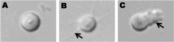
Fertilization results from the fusion of male and female gametes in all sexually reproducing organisms. Much of nematode fertility work was focused on Caenorhabditis elegans and Ascaris suum. The C. elegans hermaphrodite produces a limited number of sperm initially and then commits to the exclusive production of oocytes. The postmeiotic differentiation called spermiogenesis converts sessile spermatids into motile spermatozoa. The motility of spermatozoa depends on dynamic assembly and disassembly of a major sperm protein-based cytoskeleton uniquely found in nematodes. Both self-derived and male-derived spermatozoa are stored in spermatheca, the site of fertilization in hermaphrodites. The oocyte is arrested in meiotic prophase I until a sperm-derived signal relieves the inhibition allowing the meiotic maturation to occur. Oocyte undergoes meiotic maturation, enters into spermatheca, gets fertilized, completes meiosis, and exits into uterus as a zygote. This review focuses on our current understanding of the events around fertilization in nematodes.


Figures

Figure 1
Figure 1
DIC images of spermatid (A), partially activated spermatid showing spikes (arrow indicates spike/filopodium in B) and fully activated spermatozoa (arrow indicates pseudopod in C).
Figure 2
Figure 2
Schematic diagram depicting the motility machinery of Ascaris sperm. (Top) Cartoon of spermatozoan shows the in vivo location of the proteins participating in the assembly and disassembly of MSP fibers. The outer and inner leaflets of plasma membrane are represented as blue line and violet line, respectively. (Bottom) The MSP dimer (grey) constitutes the building block of MSP fiber. The MSP fiber grown in vitro is composed of two parts: vesicular part and fibrous part. Note that the plasma membrane of the vesicle that supports the assembly of MSP fiber is turned `inside out'. Unknown tyrosin kinase activates MPOP at the leading edge. Phospho-MPOP recruits MPAK which in turn phosphorylates MFP2. Phospho-MFP2 increases the rate of MSP fiber assembly. Unknown kinase phosphorylates cytosolic protein, MFP3 at multiple sites. Phospho-MFP3 binds with MFP3 to stabilize MSP fibers. The Ser/Thr phosphatase, PP2A is localized near the cell body which gets activated upon dephosphorylation by unknown phosphatase. Active PP2A dephosphorylates phospho-MFP3 and dephosphorylated MFP3 detaches from MSP fibers, thereby favoring the disassembly of MSP fibers near the cell body. Phospho group appended to the proteins are depicted as small red circle.
Figure 3
Figure 3
Cartoon depicting the pathway of sperm attractant production in C. elegans Dietary Mono Unsaturated Fatty Acids (MUFA) are converted into Poly Unsaturated Fatty Acid (PUFA) through a fat metabolic pathway in which FAT-2 plays an important role. PUFA(Red dots) are incorporated into yolk (yellow circle). The yolk enters into psedocoelom and reaches the gonad, where LDL receptor RME-2 mediates endocytosis of yolk. PUFAs are further metamolised to form a `sperm attractant' (Red stars) which promotes the directional migration of sperm to spermatheca.
Figure 4
Figure 4
Proteins complexes present at the cell cortex of oocyte required for oocyte-to-embryo transition. EGG-3 binds with CHS-1, EGG-4/5 and MBK-2. The phospho group appended to the MBK-2 by itself during translation of MBK-2 is depicted as a small red circle. Pseudophospatase EGG-4/5 binds with phospho-MBK-2 and inhibits its activity in oocyte.

Similar articles

Cited by

References

    1. Abbas M, Cain GD. In vitro activation and behavior of the ameboid sperm of Ascaris suum (Nematoda) Cell Tissue Res. 1979;200:273–84. - PubMed
    1. Argon Y, Ward S. Caenorhabditis elegans fertilization-defective mutants with abnormal sperm. Genetics. 1980;96:413–33. - PMC - PubMed
    1. Bae YK, Kim E, L'Hernault S W, Barr MM. The CIL-1 PI 5-phosphatase localizes TRP Polycystins to cilia and activates sperm in C. elegans. Curr Biol. 2009;19:1599–607. - PMC - PubMed
    1. Baker AM, Roberts TM, Stewart M. 2.6 A resolution crystal structure of helices of the motile major sperm protein (MSP) of Caenorhabditis elegans. J Mol Biol. 2002;319:491–9. - PubMed
    1. Brattig NW, Schwohl A, Rickert R, Buttner DW. The filarial parasite Onchocerca volvulus generates the lipid mediator prostaglandin E(2) Microbes Infect. 2006;8:873–9. - PubMed

Publication types

LinkOut - more resources