Review
. 2011 Nov;1811(11):838-53.
doi: 10.1016/j.bbalip.2011.06.027. Epub 2011 Jul 1.

Analysis of mammalian sphingolipids by liquid chromatography tandem mass spectrometry (LC-MS/MS) and tissue imaging mass spectrometry (TIMS)

Affiliations
Review

Analysis of mammalian sphingolipids by liquid chromatography tandem mass spectrometry (LC-MS/MS) and tissue imaging mass spectrometry (TIMS)

M Cameron Sullards et al. Biochim Biophys Acta. 2011 Nov.

Abstract

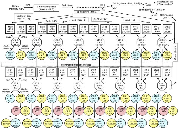
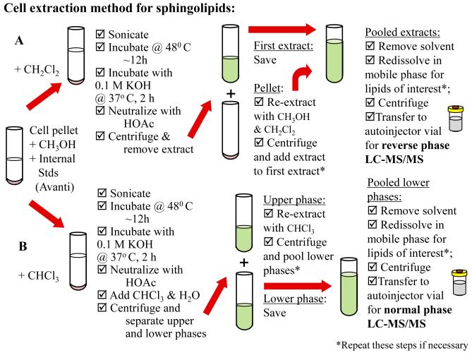
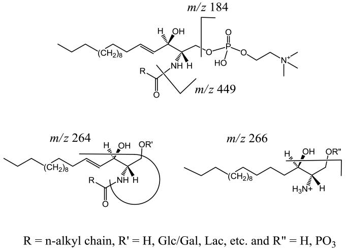
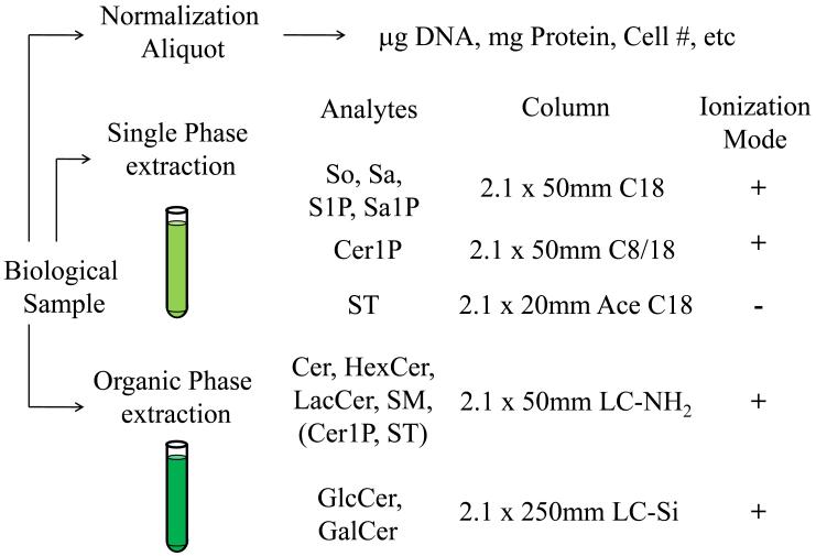
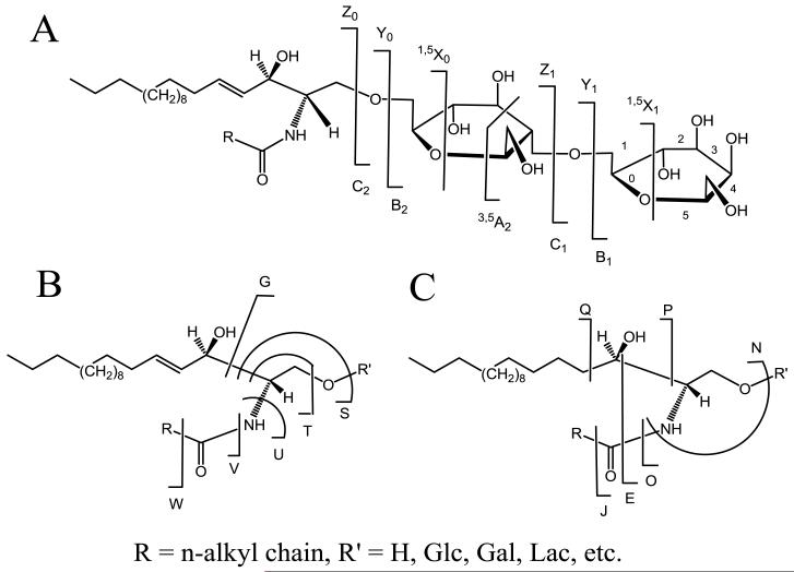
Sphingolipids are a highly diverse category of molecules that serve not only as components of biological structures but also as regulators of numerous cell functions. Because so many of the structural features of sphingolipids give rise to their biological activity, there is a need for comprehensive or "sphingolipidomic" methods for identification and quantitation of as many individual subspecies as possible. This review defines sphingolipids as a class, briefly discusses classical methods for their analysis, and focuses primarily on liquid chromatography tandem mass spectrometry (LC-MS/MS) and tissue imaging mass spectrometry (TIMS). Recently, a set of evolving and expanding methods have been developed and rigorously validated for the extraction, identification, separation, and quantitation of sphingolipids by LC-MS/MS. Quantitation of these biomolecules is made possible via the use of an internal standard cocktail. The compounds that can be readily analyzed are free long-chain (sphingoid) bases, sphingoid base 1-phosphates, and more complex species such as ceramides, ceramide 1-phosphates, sphingomyelins, mono- and di-hexosylceramides, sulfatides, and novel compounds such as the 1-deoxy- and 1-(deoxymethyl)-sphingoid bases and their N-acyl-derivatives. These methods can be altered slightly to separate and quantitate isomeric species such as glucosyl/galactosylceramide. Because these techniques require the extraction of sphingolipids from their native environment, any information regarding their localization in histological slices is lost. Therefore, this review also describes methods for TIMS. This technique has been shown to be a powerful tool to determine the localization of individual molecular species of sphingolipids directly from tissue slices.


Figures

Figure 1
Figure 1
Biosynthetic pathway of sphingolipids consisting of free long chain bases (top row), which are N-acylated by several CerS enzymes to form dihydroceramides (upper row of octagons). These DHCer may then processed by dihydroceramide desaturases to introduce a 4, 5 double bond to form ceramide (lower row of octagons). Collectively both the DHCer and Cer may be converted to more complex via addition of a polar headgroup at the 1′-OH position via sphingomyelin synthase to form sphingomyelin (thick lined squares), or ceramide kinase to form ceramide-1-phosphate (thin lined squares). Carbohydrates may also be linked to this position as well via glucosylceramide or galactosylceramide synthase to form glucosylceramides (light blue circles) and galactosylceramides (yellow circles), respectively. The former may have additional carbohydrates such as galactose complexed to it to form lactosylceramide (bottom row of light blue and yellow linked circles). The latter may also be sulfated to form the sulfatide species (pink circles). The far right and upper left corners show the degradative pathway of sphingolipids via ceramidases, sphingosine/sphinganine kinases, and sphingosine/sphinganine phosphate lyases, which constitutes the only known exit from the sphingolipid metabolic pathway.
Figure 2
Figure 2
Protocol for sphingolipidomic analyses via LC-MS/MS using (A) the single phase extraction for high recovery of LCB, their phosphates, Cer1P, and ST; and (B) the organic phase extraction for high recovery of complex sphingolipids Cer, Glc/GalCer, LacCer, and SM.
Figure 3
Figure 3
Common fragmentations of sphingomyelin (top center) showing cleave of the phosphocholine headgroup in the positive mode and neutral loss of the fatty acid in the negative mode. Complex sphingolipids fragment via neutral loss of the fatty acid and dehydration of the sphingoid base (lower left), as do the long chain sphingoid bases (lower right). These unique species are used for determination of sphingoid base, fatty acid, and head group combinations for building a “parts list” for LC-MS/MS.
Figure 4
Figure 4
The sphingolipid analysis pipeline detailing the sample normalization protocol and which classes of sphingolipids may be analyzed using a specific extraction procedure, HPLC column, and ionization mode.
Figure 5
Figure 5
A detailed nomenclature for glycosphingolipids corresponding to the (A) glycan, (B) fatty acid, and (C) sphingoid base fragment ions.

Similar articles

Cited by

References

    1. Lahiri S, Futerman AH. The metabolism and function of sphingolipids and glycosphingolipids. Cell Mol. Life Sci. 2007;64:2270–2284. - PMC - PubMed
    1. Merrill AH, Jr., Wang MD, Park M, Sullards MC. (Glyco)sphingolipidology: an amazing challenge and opportunity for systems biology. Trends Biochem. Sci. 2007;32:457–468. - PubMed
    1. Haynes CA, Allegood JC, Park H, Sullards MC. Sphingolipidomics: Methods for the Comprehensive Analysis of Sphingolipids. J. Chromatogr. B Analyt. Technol. Biomed. Life Sci. 2009;877(26):2696–2708. - PMC - PubMed
    1. Shaner RL, Allegood JC, Park H, Wang E, Kelly S, Haynes CA, Sullards MC, Merrill AH., Jr. Quantitative analysis of sphingolipids for lipidomics using triple quadrupole and quadrupole linear ion trap mass spectrometers. J. Lipid Res. 2009;50:1692–1707. - PMC - PubMed
    1. Sullards MC, Allegood JC, Kelly S, Wang E, Haynes CA, Park H, Chen Y, Merrill AH., Jr. Structure-specific, quantitative methods for analysis of sphingolipids by liquid chromatography-tandem mass spectrometry: “inside-out” sphingolipidomics. Methods Enzymol. 2007;432:83–115. - PubMed

Publication types

LinkOut - more resources