. 2012 Jul;61(7):1085-94.
doi: 10.1136/gut.2010.236026. Epub 2011 Jul 11.

Genetic evolution of pancreatic cancer: lessons learnt from the pancreatic cancer genome sequencing project

Affiliations

Genetic evolution of pancreatic cancer: lessons learnt from the pancreatic cancer genome sequencing project

Christine A Iacobuzio-Donahue. Gut. 2012 Jul.

Abstract

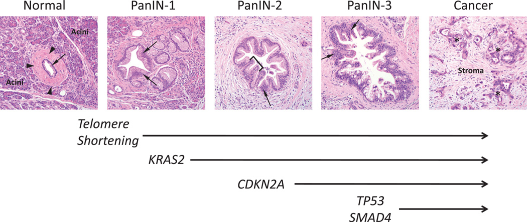
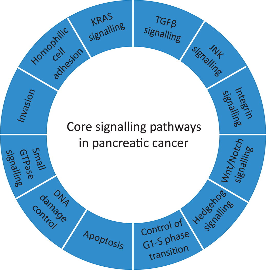
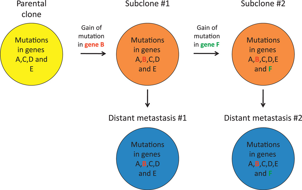
Pancreatic cancer is a disease caused by the accumulation of genetic alterations in specific genes. Elucidation of the human genome sequence, in conjunction with technical advances in the ability to perform whole exome sequencing, have provided new insight into the mutational spectra characteristic of this lethal tumour type. Most recently, exomic sequencing has been used to clarify the clonal evolution of pancreatic cancer as well as provide time estimates of pancreatic carcinogenesis, indicating that a long window of opportunity may exist for early detection of this disease while in the curative stage. Moving forward, these mutational analyses indicate potential targets for personalised diagnostic and therapeutic intervention as well as the optimal timing for intervention based on the natural history of pancreatic carcinogenesis and progression.


Conflict of interest statement

There are no relevant financial conflicts to disclose.

Figures

Figure 1
Figure 1
Morphological and genetic progression model of pancreatic carcinogenesis. Histological examples of a normal pancreatic duct, pancreatic intraepithelial neoplasia (PanIN) and pancreatic cancer are shown. Normal ducts are characterised by a low cuboidal epithelium (arrow) surrounded by a periductal fibrotic cuff (arrowheads). Ductal epithelium is relatively sparse compared with the surrounding acinar component. PanIN-1 lesions are differentiated from normal ductal epithelium by the presence of mucinous hyperplasia of the ductal cells (arrows) but without cytological atypia. By contrast, PanIN-2 lesions are notable for the presence of nuclear enlargement, atypia, crowding (arrow) and papillary infoldings of the epithelium (brackets). PanIN-3 lesions, synonymous to high-grade dysplasia/carcinoma in situ, show a complete loss of cell polarity (arrows) and marked cytological atypia in association with frequent mitotic figures and pseudopapillary growth of the neoplastic epithelium. PanIN-3 lesions may progress to invasive cancer, characterised by poorly formed neoplastic glands (asterisk) with an infiltrative growth pattern. Note the abundant desmoplastic stroma that is also a common feature of pancreatic cancer. Based on this progression model, the molecular alterations that accumulate during pancreatic carcinogenesis can be classified into early (telomere shortening and activating mutations in KRAS2), intermediate (inactivating mutations or epigenetic silencing of p16/CDKN2A) and late (inactivating mutations of TP53 and SMAD4) events. Mutations in additional genes may also occur during PanIN formation but are not illustrated in this example.
Figure 2
Figure 2
Cystic precursors of pancreatic cancer. (A) Low power view of an intraductal papillary mucinous neoplasm. This precursor develops from ductal cells lining the main pancreatic duct (arrow) leading to cystic dilation and obstruction of the main pancreatic duct by the papillary neoplasm. (B) Low power view of a mucinous cystadenoma. Unlike IPMNs that arise within the main pancreatic duct, mucinous cystadenomas are not associated with the pancreatic duct system. Note the normal pancreatic tissue in the upper right of the image that is distinct from the cystic neoplasm. In this example the neoplasm also contains foci of borderline to high-grade dysplasia (indicated by arrow). (C) Higher power view of the cystic neoplasm shown in (B) to illustrate the presence of ovarian-like stroma that underlies the mucinous epithelium. IPMN, intraductal papillary mucinous neoplasm; MCN, mucinous cystadenoma.
Figure 3
Figure 3
Core signalling pathways in pancreatic cancer. The 12 pathways and processes whose component genes were genetically altered in most pancreatic cancers based on whole exome sequencing are shown. Therapeutic targeting of one or more of these pathways, rather than specific gene alterations that occur within a pathway, provides a new paradigm for treatment of this disease. GTPase, guanosine triphosphatase; TGFβ, transforming growth factor β.
Figure 4
Figure 4
Comparative lesion sequencing. Comparative lesion sequencing (CSL) requires at least two geographically or temporally distinct samples of a patient’s cancer. These samples may be any number of synchronous metastases, the primary carcinoma and a subsequent metastatic recurrence, or even samples taken from different regions of a single primary carcinoma. In the first step of CSL, one sample (the ‘index’ lesion) is analysed by whole exome sequencing to identify all somatic mutations present in the coding fraction of the genome. In the second step of CSL, typically performed using standard sequencing methods, the presence or absence of all mutations found in the index lesion is assessed in additional samples for that patient. The ‘relatedness’ of the different samples to each other can then be derived based on the number of mutations shared. For example, mutations common to all samples are called founder mutations and reflect the genetic features of the clonal population (the ‘parental’ clone) that gave rise to all the samples analysed. By contrast, those mutations that are only present in a subset of the samples are called progressor mutations as they reflect clonal progression that occurred beyond formation of the parental clone. In the hypothetical example shown, whole exome sequencing is performed on one sample (the ‘index’ lesion shown in grey) that identified mutations in genes A–F. In the next step, two additional samples from this patient (shown in red) are analysed to determine if one or more of this set of six mutations were also present. A comparison of the findings indicates that mutations in genes A, C, D and E are common to all samples and thus represent founder mutations contained within the parental clone that gave birth to all three. By contrast, the mutations in genes B and F are only present in two of three samples and represent progressor mutations that occurred relatively later than the founder mutations in the clonal evolution of this carcinoma.
Figure 5
Figure 5
Clonal progression of pancreatic cancer. A representative example of the proposed clonal evolution of pancreatic cancer based on sequencing data of five different samples is shown. In this model, after development of the parental clone that seeded the infiltrating carcinoma (indicated in yellow), ongoing clonal evolution continues within the primary carcinoma leading to the development of a subclone characterised by the gain of a mutation in gene B. Additional clonal evolution beyond this subclone leads to the development of a second subclone characterised by the presence of a mutation in gene F. Subclones may then seed metastases to distant sites (indicated in blue) as reflected by the identical pattern of mutations in subclone 1 compared with distant metastasis 1, and in subclone 2 compared with distant metastasis 2.
Figure 6
Figure 6
Estimates of time taken for the genetic progression of pancreatic cancer. Pancreatic carcinogenesis begins with an initiating mutation in a normal cell that confers a selective growth advantage. Successive waves of clonal expansion occur in association with the acquisition of additional mutations, corresponding to the progression model of pancreatic intraepithelial neoplasia (PanIN) and time T1. One founder cell within a PanIN lesion will seed the parental clone and hence initiate the infiltrating carcinoma. This is the beginning of time T2. Following additional waves of clonal expansion, the subclones that will give rise to one or more distant metastases will develop within the infiltrating carcinoma signifying the beginning of time T3. The estimated average time for each interval is also indicated and corresponds to a total of 21.2 years from tumour initiation until the patient’s death from metastatic disease. Unfortunately, most patients are not diagnosed until late in time T3 when the cells of these metastatic subclones have already escaped the pancreas and started to grow within distant organs.

Similar articles

Cited by

References

    1. Jemal A, Siegel R, Xu J, et al. Cancer statistics, 2010. CA Cancer J clin. 2010;60:277–300. - PubMed
    1. Malvezzi M, Arfe A, Bertuccio P, et al. European cancer mortality predictions for the year 2011. Ann Oncol. 2011;22:947–956. - PubMed
    1. Maitra A, Hruban RH. Pancreatic cancer. Annu Rev Pathol. 2008;3:157–188. - PMC - PubMed
    1. Moskaluk CA, Hruban RH, Kern SE. p16 and K-ras gene mutations in the intraductal precursors of human pancreatic adenocarcinoma. Cancer Res. 1997;57:2140–2143. - PubMed
    1. Wilentz RE, Iacobuzio-Donahue CA, Argani P, et al. Loss of expression of Dpc4 in pancreatic intraepithelial neoplasia: evidence that DPC4 inactivation occurs late in neoplastic progression. Cancer Res. 2000;60:2002–2006. - PubMed

Publication types