. 2011 Nov;10(11):M111.009506.
doi: 10.1074/mcp.M111.009506. Epub 2011 Jul 11.

S-bacillithiolation protects against hypochlorite stress in Bacillus subtilis as revealed by transcriptomics and redox proteomics

Affiliations

S-bacillithiolation protects against hypochlorite stress in Bacillus subtilis as revealed by transcriptomics and redox proteomics

Bui Khanh Chi et al. Mol Cell Proteomics. 2011 Nov.

Abstract

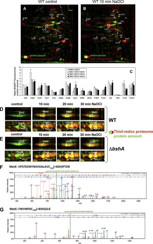
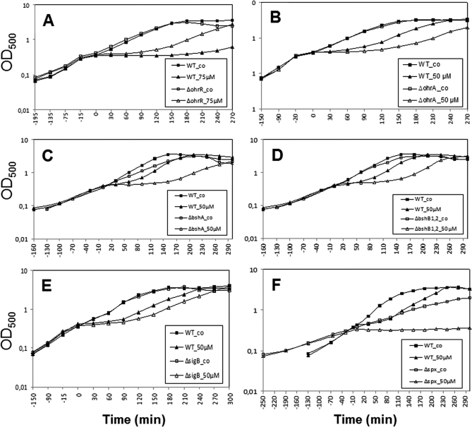
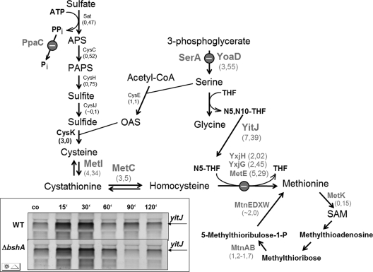
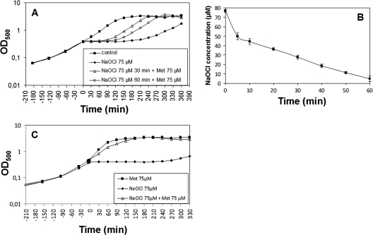
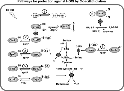
Protein S-thiolation is a post-translational thiol-modification that controls redox-sensing transcription factors and protects active site cysteine residues against irreversible oxidation. In Bacillus subtilis the MarR-type repressor OhrR was shown to sense organic hydroperoxides via formation of mixed disulfides with the redox buffer bacillithiol (Cys-GlcN-Malate, BSH), termed as S-bacillithiolation. Here we have studied changes in the transcriptome and redox proteome caused by the strong oxidant hypochloric acid in B. subtilis. The expression profile of NaOCl stress is indicative of disulfide stress as shown by the induction of the thiol- and oxidative stress-specific Spx, CtsR, and PerR regulons. Thiol redox proteomics identified only few cytoplasmic proteins with reversible thiol-oxidations in response to NaOCl stress that include GapA and MetE. Shotgun-liquid chromatography-tandem MS analyses revealed that GapA, Spx, and PerR are oxidized to intramolecular disulfides by NaOCl stress. Furthermore, we identified six S-bacillithiolated proteins in NaOCl-treated cells, including the OhrR repressor, two methionine synthases MetE and YxjG, the inorganic pyrophosphatase PpaC, the 3-D-phosphoglycerate dehydrogenase SerA, and the putative bacilliredoxin YphP. S-bacillithiolation of the OhrR repressor leads to up-regulation of the OhrA peroxiredoxin that confers together with BSH specific protection against NaOCl. S-bacillithiolation of MetE, YxjG, PpaC and SerA causes hypochlorite-induced methionine starvation as supported by the induction of the S-box regulon. The mechanism of S-glutathionylation of MetE has been described in Escherichia coli also leading to enzyme inactivation and methionine auxotrophy. In summary, our studies discover an important role of the bacillithiol redox buffer in protection against hypochloric acid by S-bacillithiolation of the redox-sensing regulator OhrR and of four enzymes of the methionine biosynthesis pathway.


Figures

Fig. 1.
Fig. 1.
Growth curves in response to sub-lethal concentrations of NaOCl stress in B. subtilis. B. subtilis wild type was grown in minimal medium to an OD500 of 0.4 and exposed to 50, 75, and 100 μm of NaOCl indicated by time point zero.
Fig. 2.
Fig. 2.
Transcriptome changes of NaOCl-induced regulons (CtsR, Spx, PerR, OhrR, ArsR, CzrA, SigmaD, and SigmaB) and of the pstSCAB operon. Fold-changes are average induction ratios of genes induced in NaOCl-treated cells versus untreated cells calculated from three transcriptome replicates with standard deviations given as errors bars. Shown are only representative genes of each regulon that are more than fivefold induced by NaOCl in supplemental Tables S1 and S3.
Fig. 3.
Fig. 3.
Northern blot analysis of selected thiol-stress specific genes of the Spx, CymR, S-box, PerR, OhrR, and MarR/DUF24 regulons in response to NaOCl, diamide, and CHP stress. Transcript analysis of nfrA, trxA, and katA indicates the induction of the Spx and PerR-regulons by NaOCl and diamide stress. The CymR-controlled cysK gene is strongly induced by diamide. The S-box regulon gene yitJ responds most strongly to NaOCl stress. The ohrA gene is controlled by the OhrR repressor and strongly induced by NaOCl and CHP. The YodB and CatR-controlled azoR1 and catDE genes are strongly up-regulated by diamide. Cells were grown to an OD500 of 0.4 and harvested before (control conditions, co) and 10 min after exposure to 50 μm NaOCl, 100 μm CHP, or 1 mm diamide. The RNA isolation and Northern blot hybridization was performed as described in the Methods section. The arrows point toward the sizes of the specific transcripts.
Fig. 4.
Fig. 4.
Hierarchical clustering analysis of RES and NaOCl induced gene expression profiles. Log2-fold changes of gene expression ratios were clustered for 1 mm diamide (Dia), 2.4 mm catechol (Cat), 0.5 mm methylhydroquinone (MHQ), 1 mm formaldehyde, 2.8 mm and 5.6 mm methylglyoxal (MG-2 and MG-5), and 50 μm NaOCl stress using the Treeview software. The cluster analysis resulted in 14 different nodes that are enriched for RES and NaOCl-induced regulons (CtsR, Spx, ArsR, CzrA, CsoR, SigmaD), the RES-specific CymR regulon, quinone-responsive regulons (PerR, YodB, CatR (YvaP), MhqR), aldehyde-responsive regulons (HxlR, AdhR, LexA) and NaOCl-specific regulons (SigmaB, OhrR). For the cluster analysis 630 genes were selected as listed in supplemental Table S4 that are induced by RES and NaOCl stress. Red indicates induction and green repression under the stress specific conditions.
Fig. 5.
Fig. 5.
ABC. The thiol-redox proteome (red) in comparison to the protein amount image (green) at control conditions (A) and after exposure to 50 μm NaOCl (B) in the wild type. Reduced protein thiols in cell extracts were alkylated with IAM followed by reduction of oxidized protein thiols with TCEP and labeling with BODIPY FL C1-IA. Proteins with reversible thiol-modifications in the control and NaOCl redox proteome are labeled in white and newly oxidized proteins in NaOCl-treated cells are labeled in red. The oxidized proteins were identified by MALDI-TOF-TOF MS/MS as shown in detail in supplemental Fig. S1A–P. C, The fluorescence/protein amount ratios are quantified as oxidation ratios at control conditions (control) and 10, 20, and 30 min after exposure to 50 μm NaOCl stress as shown in the diagram. DEFG. Close-ups of the main NaOCl-sensitive proteins MetE and GapA in the thiol-redox proteome of the wild type (D) and bshA mutant (E) and CID MS/MS spectra of the S-bacillithiolated Cys719 and Cys730 peptides of MetE (F, G). Figs. D and E show sections of reversibly oxidized proteins after NaOCl stress in the thiol-redox proteome (red) in comparison to the protein amount image (green) at control conditions (co) and 10, 20, and 30 min after exposure to 50 μm NaOCl. Figs. F and G show the CID MS/MS spectra of the S-bacillithiolated Cys719-and Cys730-peptides of MetE identified in the wild-type proteome using LTQ-Orbitrap-Velos mass spectrometry as described in the Methods section. The MS/MS spectra show the characteristic abundant precursor ions that have lost malate indicated by parent-134 (HOOC-CH2-CHOH-COOH). The Xcorr and ΔCn scores and peptide masses are listed in Table II and the corresponding b and y fragment ion series for the modified peptides are given in detail in supplemental Fig. S3B.
Fig. 6.
Fig. 6.
The OhrA peroxiredoxin and BSH protect cells against NaOCl toxicity. Growth phenotype of B. subtilis wild type (wt) in comparison to the ΔohrR (A), ΔohrA (B), ΔbshA (C), ΔbshB1,B2 (D), ΔsigB (E), and Δspx (F) mutant strains that were treated with 50 or 75 μm NaOCl at an OD500 of 0.4.
Fig. 7.
Fig. 7.
Pathways for sulfur assimilation, cysteine and methionine biosynthesis, and induction of S-box regulon genes in response to NaOCl stress. The S-box regulon gene products that are induced in the transcriptome as a result of methionine starvation provoked by the S-bacillithiolation of MetE and YxjG are bold-faced in gray. The transcriptome induction ratios are shown below the gene products. The gene products of the sulfate assimilation pathway (Sat, CysC, CysH, CysJI, CysE) are not induced upon NaOCl stress (except for CysK). The inset shows the transcriptional induction of yitJ in the wild type and ΔbshA mutant using Northern blot analysis before (co) and at different times (10, 30, 60, 90, and 120 min) after exposure to 50 μm NaOCl stress. Abbreviations: APS, adenosine-5′-phosphosulfate; PAPS, 3′-phosphoadenosine-5′-phosphosulfate; OAS, O-acetylserine; N5,N10-THF, 5,10-methylenetetrahydrofolate; N5 -THF, 5-methyltetrahydrofolate; THF, tetrahydrofolate. This figure was adapted from Tomsic et al., 2008 (52).
Fig. 8.
Fig. 8.
NaOCl stress causes methionine auxotrophy that is abolished by methionine addition. Growth phenotypes of the B. subtilis wild type treated with 75 μm NaOCl at an OD500 of 0.4. A, Methionine was added 30 or 60 min after exposure to 75 μm NaOCl stress and the growth was resumed. B, The concentrations of consumed NaOCl of wild-type cells were monitored in the culture supernatants after exposure to 75 μm NaOCl using the FOX assay. The NaOCl concentrations are given as mean values of three independent experiments with error bars. C, Methionine (75 μm) was added after inoculation to the culture at an OD500 of 0,07 and 75 μm NaOCl was added when cells had reached an OD500 of 0.4.
Fig. 9.
Fig. 9.
Proposed defensive mechanisms against hypochloric acid stress in B. subtilis. Exposure of B. subtilis to NaOCl induces an oxidative, disulfide and general stress response (OhrR, Spx, CtsR, PerR, SigmaB). NaOCl leads to S-bacillithiolation of OhrR, MetE, YxjG, PpaC, SerA, and YphP and to intramolecular disulfide formation in GapA. The thiol-disulfide isomerase YphP could function as bacilliredoxin in reduction of S-bacillithiolated proteins. (1) The redox buffer BSH could be directly involved in NaOCl detoxification leading to BSSB formation. (2) S-bacillithiolation of OhrR causes induction of the OhrA peroxiredoxin that is involved in specific NaOCl detoxification. (3) S-bacillithiolation of the inorganic pyrophosphatase PpaC could lead to decreased ATP sulfurylase activity as the removal of PPi is prevented. (4) S-bacillithiolation of the phosphoglycerate dehydrogenase SerA causes decreased levels of serine that is required for cysteine and methionine biosynthesis. (5) S-bacillithiolation of the active site Cys residues of MetE and YxjG leads to methionine auxotrophy to stop translation during the time of NaOCl detoxification (6) The glyceraldehyde-3-phosphate dehydrogenase GapA is inhibited causing decreased glycolysis. The stars indicate the redox-sensing Cys residues that are S-bacillithiolated in OhrR (Cys15), MetE (Cys730), YxjG (Cys346), or oxidized to an intramolecular disulfide in GapA (Cys152). Abbreviations: APS, adenosine-5′-phosphosulfate; N5-THF, 5-methyltetrahydrofolate; THF, tetrahydrofolate; 3-PG, 3-d-phosphoglycerate, GA-3-P, glyceraldehyde-3-phosphate; 1,3-BPG, 1,3-Bisphosphoglycerate.

Similar articles

Cited by

References

    1. Imlay J. A. (2003) Pathways of oxidative damage. Annu. Rev. Microbiol. 57, 395–418 - PubMed
    1. Imlay J. A. (2008) Cellular defenses against superoxide and hydrogen peroxide. Annu. Rev. Biochem. 77, 755–776 - PMC - PubMed
    1. Davies M. J. (2011) Myeloperoxidase-derived oxidation: mechanisms of biological damage and its prevention. J. Clin. Biochem. Nutr. 48, 8–19 - PMC - PubMed
    1. Hawkins C. L., Pattison D. I., Davies M. J. (2003) Hypochlorite-induced oxidation of amino acids, peptides and proteins. Amino Acids 25, 259–274 - PubMed
    1. Faulkner M. J., Helmann J. D. (2011) Peroxide stress elicits adaptive changes in bacterial metal ion homeostasis. Antioxid Redox Signal. doi 10.1089/ars.2010.3682 - DOI - PMC - PubMed

Publication types

MeSH terms