. 2011 Aug;138(15):3213-24.
doi: 10.1242/dev.058693.

The L6 domain tetraspanin Tm4sf4 regulates endocrine pancreas differentiation and directed cell migration

Affiliations

The L6 domain tetraspanin Tm4sf4 regulates endocrine pancreas differentiation and directed cell migration

Keith R Anderson et al. Development. 2011 Aug.

Abstract

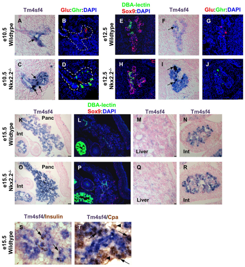
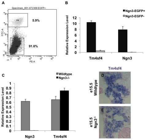
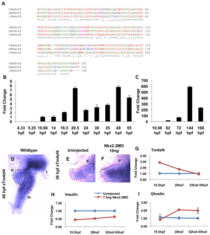
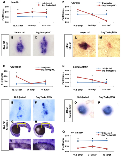
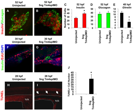
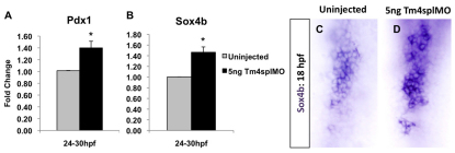
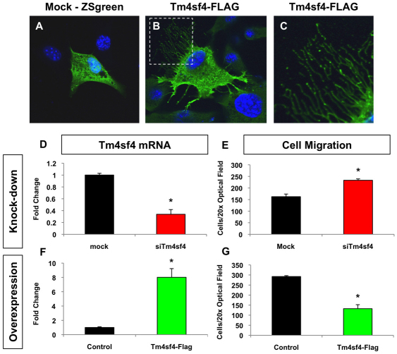
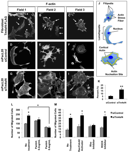
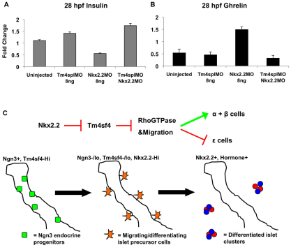
The homeodomain transcription factor Nkx2.2 is essential for pancreatic development and islet cell type differentiation. We have identified Tm4sf4, an L6 domain tetraspanin family member, as a transcriptional target of Nkx2.2 that is greatly upregulated during pancreas development in Nkx2.2(-/-) mice. Tetraspanins and L6 domain proteins recruit other membrane receptors to form active signaling centers that coordinate processes such as cell adhesion, migration and differentiation. In this study, we determined that Tm4sf4 is localized to the ductal epithelial compartment and is prominent in the Ngn3(+) islet progenitor cells. We also established that pancreatic tm4sf4 expression and regulation by Nkx2.2 is conserved during zebrafish development. Loss-of-function studies in zebrafish revealed that tm4sf4 inhibits α and β cell specification, but is necessary for ε cell fates. Thus, Tm4sf4 functional output opposes that of Nkx2.2. Further investigation of how Tm4sf4 functions at the cellular level in vitro showed that Tm4sf4 inhibits Rho-activated cell migration and actin organization in a ROCK-independent fashion. We propose that the primary role of Nkx2.2 is to inhibit Tm4sf4 in endocrine progenitor cells, allowing for delamination, migration and/or appropriate cell fate decisions. Identification of a role for Tm4sf4 during endocrine differentiation provides insight into islet progenitor cell behaviors and potential targetable regenerative mechanisms.


Figures

Fig. 1.
Fig. 1.
Tm4sf4 is expressed in the early mouse pancreatic epithelium and is upregulated in Nkx2.2–/– pancreata. (A-T) Pancreatic Tm4sf4 expression was assessed by in situ hybridization at e10.5 (A,C), e12.5 (F,I) and e15.5 (K,O,S,T) in both wild-type and Nkx2.2–/– mice. Colocalization of Tm4sf4 with glucagon (red), Sox9 (red), lectin (green), ghrelin (green), insulin (brown) and CpA (brown) was analyzed on adjacent sections (B,D,E,G,H,J,L,P,S,T). Examples of Tm4sf4HI regions are identified by arrows. Dashed lines in B and D encircle the pancreatic epithelium. Tm4sf4 expression was analyzed in the liver (M,Q) and intestine (N,R) of wild-type and Nkx2.2–/– mice. All images are 20× magnification, except K,L,O,P which are 10× and S,T which are 40×. Int, intestine; Panc, pancreas.
Fig. 2.
Fig. 2.
Mouse Ngn3+ pancreatic endocrine progenitor cells are enriched with Tm4sf4. (A) Fluorescence-activated cell sorting (FACS) plot showing the gates and percentages obtained for GFP+ and GFP populations. (B,C) qRT-PCR for Tm4sf4 and Ngn3 mRNA from Ngn3+ (black) and Ngn3 (gray) cells from FACS experiments (B), and wild-type (gray) or Ngn3–/– (black) pancreata (C). Error bars represent s.e.m. (D,E) Tm4sf4 in situ hybridization analysis on e15.5 wild-type (D) and Ngn3–/– (E) pancreata.
Fig. 3.
Fig. 3.
The tm4sf4 gene and regulation by nkx2.2a is conserved in zebrafish. (A) CLUSTAL W species alignment between human, mouse and zebrafish Tm4sf4. Identical (*), highly conserved (:) and weakly conserved (.) amino acids are indicated. Small/hydrophobic residues (red), acidic residues (blue), basic residues (magenta), hydroxyl/amine/basic residues (green) are shown. (B,C) Temporal qRT-PCR tm4sf4 mRNA analysis of early (B) and late (C) stage zebrafish embryos. (D-F) tm4sf4 in situ hybridization on wild-type (D,E) and nkx2.2a morphant (F) embryos. In, intestine; L, liver; P, pancreas. Images taken at 20× magnification. (G-I) qRT-PCR comparing uninjected (blue) and nkx2.2a morphant (red) embryo expression of tm4sf4 (G), insulin (H) and ghrelin (I). Error bars represent s.e.m.
Fig. 4.
Fig. 4.
tm4sf4 inhibits α and β cell specification, and is necessary for ε cell fates. (A-P) Hormone mRNA levels were measured by qRT-PCR comparing uninjected (blue) and tm4sf4 morphant (red) zebrafish embryos: insulin (A), glucagon (D), ghrelin (K), somatostatin (N). Hormone mRNA expression pattern was determined by in situ hybridization comparing uninjected and tm4sf4 morphant embryos at 20.5 hpf for insulin (B,C) and glucagon (E-J), and at 48 hpf for ghrelin (L,M) and somatostatin (O,P). All images were taken at 40× magnification except G,H taken at 10×; yellow boxes represent areas enlarged in I and J. (Q) Knockdown efficiency of Tm4splMO was determined at all stages. Error bars represent s.e.m. *P<0.05.
Fig. 5.
Fig. 5.
β cells are increased independently of proliferation, ε cells are decreased and aberrant α cells undergo apoptosis as a consequence of tm4sf4 loss. (A,B) Immunofluorescence of insulin (red) and glucagon (green) in uninjected (A) and tm4sf4 morphant (B) zebrafish embryos at 52 hpf. Confocal images were taken at 25× magnification with 2× digital zoom. (C-E) Total β (C) and α (D) cells were counted from confocal z-stacks (n=6). Total ε cells (E) were counted from in situ hybridization 20× images (n=6). (F,G) β cell proliferation was assessed by confocal microscopy of EdU (red) incorporation and immunofluorescence for insulin (green) in uninjected (F) and tm4sf4 morphant (G) embryos. (H-J) TUNEL assay to assess apoptotic cells in the presumptive intestine of 28 hpf embryos. Arrows indicate TUNEL+ cells. Dashed line delineates the yolk. Error bars represent s.e.m. *P<0.05.
Fig. 6.
Fig. 6.
pdx1 and sox4b expression are increased in tm4sf4 morphant zebrafish embryos. (A,B) mRNA levels were measured at 24-30 hpf comparing uninjected embryos (gray) and tm4sf4 morphants (black) for pdx1 (A) and sox4b (B) by qRT-PCR. Error bars represent s.e.m. *P<0.05. (C,D) sox4b expression pattern was analyzed by in situ hybridization at 18 hpf.
Fig. 7.
Fig. 7.
Tm4sf4 alters cell morphology and inhibits cell migration. (A-C) Cell morphology was analyzed in mock/ZSgreen (A) and Tm4sf4-FLAG (B,C) transfected (green) NIH 3T3 cells. Nuclei were stained with DAPI and confocal images were acquired at 63×. C shows an enlargement of the boxed area in B. (D-G) Number of cells migrated in Boyden chamber assays compared with mock transfected cells when endogenous Tm4sf4 is knocked down in mPacL20 ductal epithelial cells (D,E) or when Tm4sf4-FLAG is overexpressed in mPacL20 cells (F,G). Error bars represent s.e.m. *P<0.05.
Fig. 8.
Fig. 8.
Tm4f4 disrupts actin organization and migration through a ROCK-independent Rho GTPase pathway. (A-I) F-actin localization and organization was analyzed by rhodamine phalloidin staining in NIH 3T3 fibroblast (A) and 293T fibroblast (B,C) cells expressing (arrows) or not expressing (arrowheads) Tm4sf4-FLAG, as well as in mPacL20 ductal epithelial cells transfected with control siRNA (D-F) or siTm4sf4 (G-I). Confocal magnification 63×. (J) Schematic depicting cytoskeletal actin structures (yellow) affected by Tm4sf4. (K) Percentage of mPacL20 cells exhibiting the phenotype displayed in G-I. (L,M) Nuclei positive cells migrated in Boyden chamber assays were compared in siControl (gray) and siTm4sf4 (black) transfected cells 16 hours after cells were treated with Rho family GTPase modulators. Error bars represent s.e.m. *P<0.05, **P<0.01.
Fig. 9.
Fig. 9.
Tm4sf4 is a critical factor downstream of Nkx2.2 and a model for a mechanism in endocrine islet cell fate specification. (A,B) Suppression analysis was performed in 28 hpf zebrafish by injecting antisense morpholinos against both tm4sf4 and nkx2.2a. Differences in uninjected, single morphant and double morphant embryos were compared for insulin (A) and ghrelin (B) mRNA by qRT-PCR. Error bars represent s.e.m. (C) Our model proposes that Tm4sf4 is highly expressed within the Ngn3+ progenitors inhibiting Rho GTPase-mediated detachment and migration. Nkx2.2 is required to downregulate Tm4sf4 and ultimately promote correct islet cell differentiation.

References

    1. Anderson K. R., Torres C. A., Solomon K., Becker T. C., Newgard C. B., Wright C. V., Hagman J., Sussel L. (2009a). Cooperative transcriptional regulation of the essential pancreatic islet gene NeuroD1 (beta2) by Nkx2.2 and neurogenin 3. J. Biol. Chem. 284, 31236-31248 - PMC - PubMed
    1. Anderson K. R., White P., Kaestner K. H., Sussel L. (2009b). Identification of known and novel pancreas genes expressed downstream of Nkx2.2 during development. BMC Dev. Biol. 9, 65 - PMC - PubMed
    1. Bard J. B., Hay E. D. (1975). The behavior of fibroblasts from the developing avian cornea. Morphology and movement in situ and in vitro. J. Cell Biol. 67, 400-418 - PMC - PubMed
    1. Biemar F., Argenton F., Schmidtke R., Epperlein S., Peers B., Driever W. (2001). Pancreas development in zebrafish: early dispersed appearance of endocrine hormone expressing cells and their convergence to form the definitive islet. Dev. Biol. 230, 189-203 - PubMed
    1. Chen W. T. (1981). Mechanism of retraction of the trailing edge during fibroblast movement. J. Cell Biol. 90, 187-200 - PMC - PubMed

Publication types

MeSH terms