. 2011 Aug;138(15):3261-72.
doi: 10.1242/dev.063602.

Generation of mice with functional inactivation of talpid3, a gene first identified in chicken

Affiliations

Generation of mice with functional inactivation of talpid3, a gene first identified in chicken

Fiona Bangs et al. Development. 2011 Aug.

Abstract

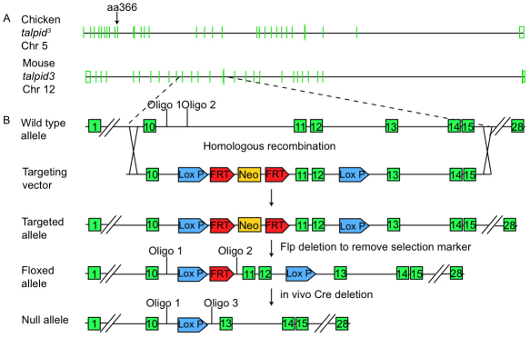
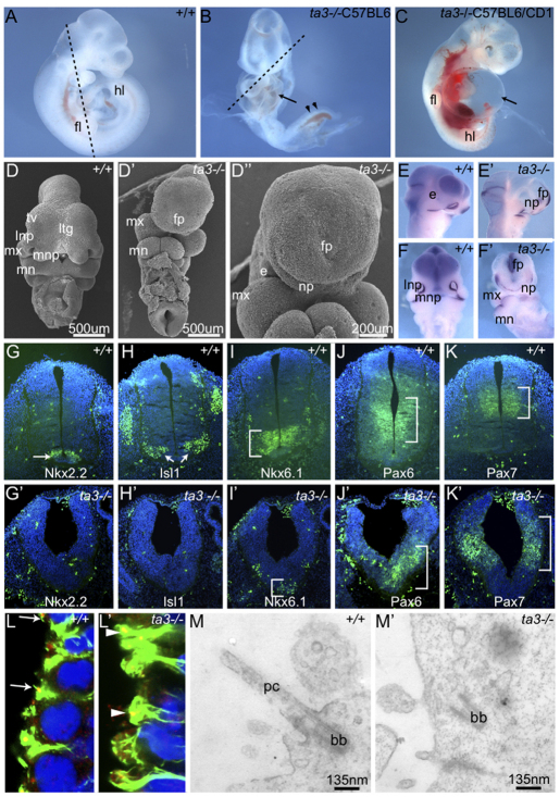
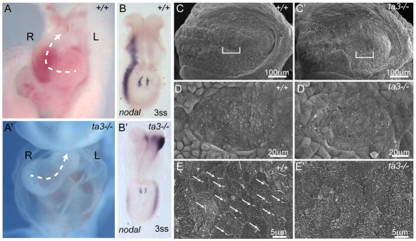
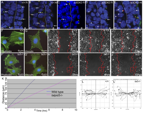
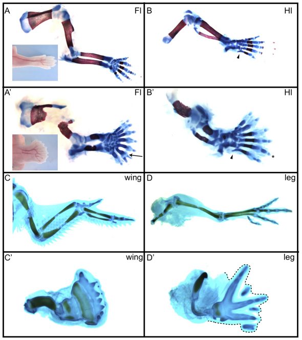
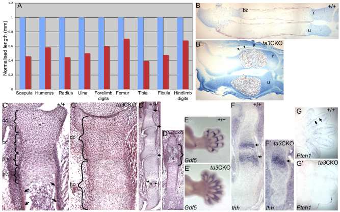
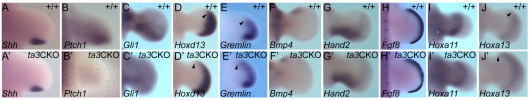
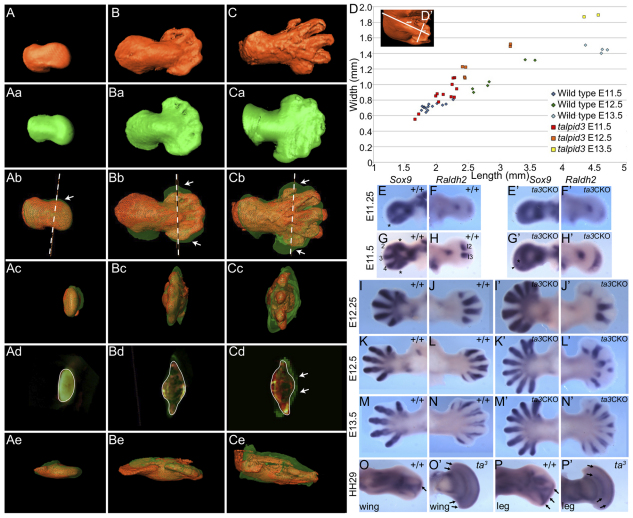
Specification of digit number and identity is central to digit pattern in vertebrate limbs. The classical talpid(3) chicken mutant has many unpatterned digits together with defects in other regions, depending on hedgehog (Hh) signalling, and exhibits embryonic lethality. The talpid(3) chicken has a mutation in KIAA0586, which encodes a centrosomal protein required for the formation of primary cilia, which are sites of vertebrate Hh signalling. The highly conserved exons 11 and 12 of KIAA0586 are essential to rescue cilia in talpid(3) chicken mutants. We constitutively deleted these two exons to make a talpid3(-/-) mouse. Mutant mouse embryos lack primary cilia and, like talpid(3) chicken embryos, have face and neural tube defects but also defects in left/right asymmetry. Conditional deletion in mouse limb mesenchyme results in polydactyly and in brachydactyly and a failure of subperisoteal bone formation, defects that are attributable to abnormal sonic hedgehog and Indian hedgehog signalling, respectively. Like talpid(3) chicken limbs, the mutant mouse limbs are syndactylous with uneven digit spacing as reflected in altered Raldh2 expression, which is normally associated with interdigital mesenchyme. Both mouse and chicken mutant limb buds are broad and short. talpid3(-/-) mouse cells migrate more slowly than wild-type mouse cells, a change in cell behaviour that possibly contributes to altered limb bud morphogenesis. This genetic mouse model will facilitate further conditional approaches, epistatic experiments and open up investigation into the function of the novel talpid3 gene using the many resources available for mice.


Figures

Fig. 1.
Fig. 1.
Structure of chicken and mouse genes and strategy for generating a talpid3–/– mouse. (A) A comparison of the chicken talpid3 gene (KIAA0586, chromosome 5) with the mouse talpid3 gene (2700049A03Rik, chromosome 12). Green bars represent exons. Arrow marks the exon that includes amino acid 366, which is mutated in chicken talpid3, forming a premature stop codon. (B) Magnified region of mouse talpid3 showing the cloning strategy for generating the talpid3–/– mouse. Shown are the loxP sites (blue) flanking exons 11 and 12 (green), FLP recognition target (FRT) sites (red) and the neomycin cassette (yellow) used as selection marker. Annealing sites of genotyping oligos are indicated.
Fig. 2.
Fig. 2.
Phenotype of talpid3–/– mouse embryos. (A-C) Mouse E10.5 wild-type (A) and talpid3–/– (B) littermates on C57BL/6 background, or talpid3–/– on C57BL/6/CD1 mixed background (C). Arrow indicates pericardial oedema; arrowheads indicate haemorrhaging. fl, forelimb; hl, hind limb. The dashed line indicates the plane of sections shown in Fig. S2 in the supplementary material. (D-F′) Scanning electron micrographs (D-D′) and whole-mount Fgf8 in situ hybridisation (E-F′) at E10.5 showing loss of midline facial structures in talpid3–/– embryos. e, eye; fp, frontal process; ltg, lamina terminalis groove; lnp, lateral nasal process; mn, mandibular process; mx, maxillary process; mnp, medial nasal process; np, nasal pit; tv, telencephalic vesicle. (G-K′) Transverse sections through E10.5 neural tube stained with antibodies to neural transcription factors (green); nuclei are counterstained with DAPI (blue). Expression of the ventral markers Nkx2.2 and Islet1 (arrows) is lost and Nkx6.1 (bracket) is reduced in talpid3–/– embryos (compare G-I with G′-I′). Expression of the dorsal markers Pax6 and Pax7 (brackets) is expanded in talpid3–/– embryos (compare J,K with J′,K′). (L,L′) Transverse section through the neural tube. In wild type (L), cilia axonemes (arrows; stained for acetylated tubulin, green) project into the lumen from centrosomes (stained for γ-tubulin, red), whereas no axonemes project from centrosomes (arrowheads) in talpid3–/– mutant cells (L′). (M,M′) Transmission electron micrographs of transverse sections though neural tube show primary cilium (pc) projecting into the lumen of a wild-type cell (M). The basal body (bb) is at the apical surface of a talpid3–/– cell but is not docked (M′).
Fig. 3.
Fig. 3.
Left/right asymmetry in wild-type and talpid3–/– mouse embryos. (A,A′) Wild-type mouse embryo heart loops to the right (A), whereas heart looping is randomised in talpid3–/– mouse embryos; an example of looping to the left is shown (A′). Dashed arrows indicate the direction of looping; R, right; L, left. (B,B′) Nodal is expressed in left lateral plate mesoderm of wild-type embryos (B), and in both the left and right lateral plate mesoderm of talpid3–/– embryos (B′). ss, somite stage. (C-E′) Scanning electron micrographs (increasing magnification top to bottom) of E8.0 node (brackets). (C-E) A primary cilium is present on almost every cell in the wild-type node (arrows). (C′-E′) Cilia are absent from the talpid3–/– node.
Fig. 4.
Fig. 4.
Loss of cilia from cells of talpid3 CKO mouse limbs. (A,B) Wild-type mouse forelimb (A) and hind limb (B). Primary cilia axonemes (arrowed; stained for acetylated tubulin, green) project from centrosomes (stained for γ-tubulin, red) on mesenchyme and ectoderm cells. (C) talpid3 CKO forelimb at E9.5, showing cilia (arrows). (D) talpid3 CKO forelimb at E10.5. Primary cilia are absent from mesenchyme cells (arrowhead shows centrosome), but present on ectoderm cells (arrows). (E) talpid3 CKO hind limb at E10.5; 2% of mesenchyme cells have primary cilium (arrow). Fl, forelimb; Hl, hind limb; m, mesenchyme and e, ectoderm, separated by dashed line. (F,F′) Phalloidin marks stress fibres (green) in wild-type mouse limb fibroblasts (F), which are reduced in talpid3–/– fibroblasts (F′); also note the increased number of filopodia around the cell circumference. (G,G′) Vinculin staining marks focal adhesions in wild-type mouse limb fibroblasts (G), which are absent in talpid3–/– fibroblasts (G′). (H-J′) Still images from film of scratch assay. Wild-type mouse limb fibroblasts migrate to close the scratch (delimited by red lines) within 10 hours (H-J), whereas talpid3–/– fibroblasts migrate more slowly (H′-J′). (K) The migration speed of wild-type mouse limb fibroblasts as compared with talpid3–/– fibroblasts. (L,L′) Plots tracing the movement of wild-type mouse limb fibroblasts (L) or talpid3–/– fibroblasts (L′) away from the scratch edge show that talpid3–/– fibroblasts lack directionality.
Fig. 5.
Fig. 5.
Skeletal patterns in wild-type and talpid3 CKO mouse limbs. (A-B′) E17.5 wild-type (A,B) and talpid3 CKO (A′,B′) mouse forelimb (Fl) and hind limb (Hl) stained with Alcian Blue for cartilage and Alizarin Red for calcified tissue. The talpid3 CKO mouse forelimb and hind limb show polydactyly. Arrow (A′) indicates bifurcated digit. Asterisk (B′) indicates penultimate posterior digit with normal morphology. Arrowheads indicate normal posterior metatarsal (compare B with B′). Insets show wild-type (in A) and talpid3 CKO (in A′) forelimbs at E17.5; note the soft tissue syndactyly in the talpid3 CKO forelimb. (C-D′) Day 11 wild-type (C,D) and talpid3 (C′,D′) chicken wing and leg stained with Alcian Blue. Note the cartilaginous syndactyly in the talpid3 chicken wing and leg (dotted line marks edge of limb).
Fig. 6.
Fig. 6.
Skeletogenesis in wild-type and talpid3 CKO mouse limbs. (A) The length of talpid3 CKO skeletal elements (red) as a percentage of wild-type elements (blue) normalised to one. (B,B′) Wild-type (B) and talpid3 CKO (B′) mouse radius (r) and ulna (u), with cartilage stained with Alcian Blue at ends of elements, and bony collar (bc) stained with Alizarin Red. In talpid3 CKO radius and ulna, cartilage is present at the ends of elements and also surrounds the Alizarin Red-stained (arrowheads) central region of calcified tissue. (C,C′) Longitudinal section through the proximal growth plate of wild-type (C) and talpid3 CKO (C′) ulna stained with Hematoxylin and Eosin. Brackets indicate different zones: dc, dividing chondrocytes; cc, columnar chondrocytes; phc, prehypertrophic chondrocytes; hc, hypertrophic chondrocytes; arrows indicate subperiosteal bone. Note only dividing and hypertrophic chondrocytes and no subperiosteal bone in talpid3 CKO ulna. (D,D′) Sections of middle digit stained with Hematoxylin and Eosin. Note defined joints (arrows), ossification (asterisk) and subperiosteal bone (arrowhead) in wild type (D), whereas the talpid3 CKO digit has an enlarged joint-forming region (bracket, D′). (E,E′) Gdf5 expression marks joints in wild-type (E) and talpid3 CKO (E′) forelimbs at E13.5. (F,F′) Longitudinal section through humerus showing Ihh expression (dark blue) in two bands (arrows) of prehypertrophic chondrocytes in wild type (F) but in only a single band (arrow) of hypertrophic like-chondrocytes in talpid3 CKO humerus (F′). (G,G′) Ptch1 is expressed (arrows) in perichondrium surrounding digits in wild-type hand plate (G) but is absent in talpid3 CKO hand plate (G′).
Fig. 7.
Fig. 7.
Gene expression in wild-type and talpid3 CKO mouse limbs. (A-J′) Whole-mount in situ hybridisation for the indicated genes in wild-type (A-J) and talpid3 CKO (A′-J′) mouse forelimb at E10.5 (A-B′) and E11.5 (C-J′). Limbs viewed from dorsal side, anterior up. Arrowheads indicate anterior extension of gene expression in talpid3–/– limb buds compared with wild type.
Fig. 8.
Fig. 8.
Limb bud shape and digit spacing in wild-type and talpid3 CKO mouse limbs. (A-Ce) Three-dimensional surfaces of right forelimbs from wild-type (A-C, red) and talpid3 CKO (Aa-Ca, green) mice. A-Ae, E11.5; B-Be, E12.5; C-Ce, E13.5. Overlaid limbs (Ab-Cb) show the difference (arrows) in shape between wild type (red mesh) and talpid3 CKO (transparent green) hand plates. Dashed line indicates the plane of sections in Ad-Cd. (Ac-Cc) Overlaid limbs viewed from a distal perspective. (Ad-Cd) Digital sections show that talpid3 CKO limbs are thickened ventrally at E13.5 (arrows). (Ae-Ce) Overlaid limbs viewed from the anterior. (D,D′) Scatter plot (D) showing increasing hand plate width over limb length (measured from limb tip to spine, see D′) in talpid3 CKO forelimbs. (E,G,I,K,M) Sox9 expression in wild-type left forelimb from E11.5 to E13.5. Digit-forming condensations are marked with an asterisk (E,G) or numbered (G). (E′,G′,I′,K′,M′) Sox9 expression in talpid3 CKO left forelimb from E11.5 to E13.5. Region with reduced Sox9 expression is marked with an asterisk (G′), and the first digit condensation is marked with an arrowhead (G′). (F,H,J,L,N) Raldh2 expression in wild-type left forelimb from E11.5 to E13.5. Interdigits are numbered (H). (F′,H′,J′,L′,N′) Raldh2 expression in talpid3 CKO left forelimb from E11.5 to E13.5. (O,P) Raldh2 expression in wild-type chicken wing (O) and leg (P). Expression is present in the interdigit and between the digit tip and apical ectodermal ridge (arrows). (O′,P′) Raldh2 expression in talpid3 chicken wing (O′) and leg (P′). Note Raldh2 expression in a thin line between metacarpals/metatarsals and phalanges as well as beneath the apical ectodermal ridge (arrows), but no interdigital Raldh2 expression. Limbs viewed from dorsal side, anterior up.

References

    1. Abbott U. K., Taylor L. W., Abplanalp H. (1959). A second talpid-like mutation in the fowl. Poult. Sci. 38, 1185
    1. Akiyama H., Chaboisssier M. C., Martin J. F., Schedl A., de Crombrugghe B. (2002). The transcription factor Sox9 has essential roles in successive steps of the chondrocyte differentiation pathway and is required for expression of Sox5 and Sox6. Genes Dev. 16, 2813-2828 - PMC - PubMed
    1. Baker K., Beales P. L. (2009). Making sense of cilia in disease: the human ciliopathies. Am. J. Med. Genet. C Semin. Med. Genet. 151C, 281-295 - PubMed
    1. Beales P. L., Elcioglu N., Woolf A. S., Parker D., Flinter F. A. (1999). New criteria for improved diagnosis of Bardet-Biedl syndrome: results of a population survey. J. Med. Genet. 36, 437-446 - PMC - PubMed
    1. Biesecker L. G. (2008). The Greig cephalopolysyndactyly syndrome. Orphanet. J. Rare Dis. 3, 10 - PMC - PubMed

Publication types

MeSH terms