. 2011 Nov;96(11):1580-8.
doi: 10.3324/haematol.2011.042515. Epub 2011 Jul 12.

Osteomyelosclerosis, anemia and extramedullary hematopoiesis in mice lacking the transcription factor NFATc2

Affiliations

Osteomyelosclerosis, anemia and extramedullary hematopoiesis in mice lacking the transcription factor NFATc2

Wolfgang Bauer et al. Haematologica. 2011 Nov.

Abstract

Background: Nuclear factors of activated T cells (NFAT) are transcription factors that are central to cytokine production in activated T cells and regulate the development and differentiation of various tissues. NFATc2 is expressed in hematopoietic stem cells and regulated during myeloid commitment in a lineage-specific manner. The biological role of NFATc2 in hematopoiesis is, however, unclear.

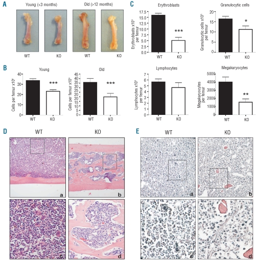
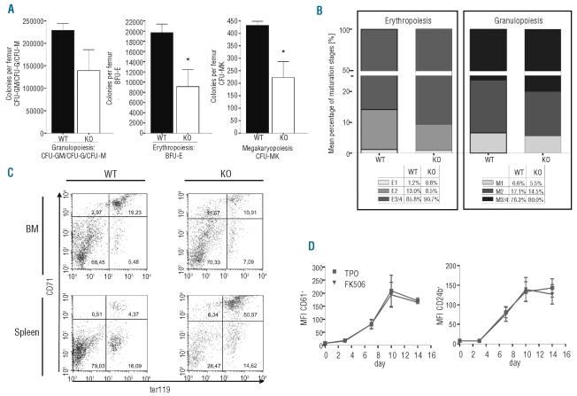
Design and methods: In the present study, we analyzed steady-state hematopoiesis in young (<3 months) and old (>12 months) mice lacking NFATc2. Complete blood counts were performed in the peripheral blood, bone marrow and spleen. Using cytological and histological analyses, the blood cell differential was determined. Colony-formation assays were used to determine the differentiation potential of hematopoietic cells. Bone cell cultures were derived from the bone marrow, and bone remodeling markers were determined in the serum.

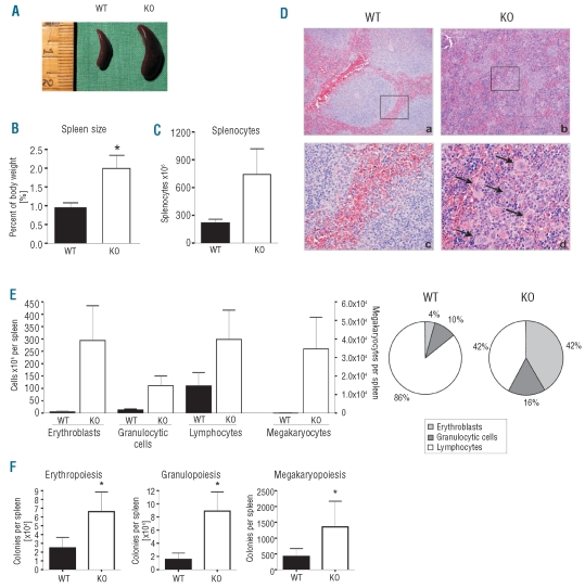
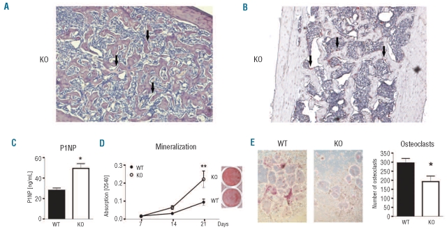
Results: NFATc2(-/-) mice older than 12 months were anemic and thrombocytopenic. The bone marrows of these mice showed a markedly reduced number of hematopoietic cells, of which megakaryocytic and erythroid lineages were most affected. While the number of hematopoietic progenitor cells in NFATc2-deficent bone marrow was reduced, the myeloid differentiation potential of these cells remained intact. Aged NFATc2(-/-) mice showed ossification of their bone marrow space and developed extramedullary hematopoiesis in the spleen. Ex vivo differentiation assays revealed an intrinsic defect of NFATc2-deficient stromal cells, in which NFATc2(-/-) osteoblasts differentiated more efficiently than wild-type cells, whereas osteoclast differentiation was impaired.

Conclusions: Our data suggest that NFATc2 may play a role in the maintenance of steady-state hematopoiesis and bone remodeling in adult organisms.


Figures

Figure 1.
Figure 1.
Bone marrow hypoplasia and osteomyelosclerosis in aged NFATc2−/− mice. (A) Femora of young and aged wild-type (WT) and NFATc2 knock-out (KO) mice. Note that femora of aged KO mice appear pale compared to WT bones. (B) Numbers of bone marrow cells extracted from young and old WT and KO femora. (C) Bone marrow differentials. The numbers of erythroblasts, granulocytic cells, lymphocytes and megakaryocytes extracted from aged WT and KO femora are shown. Results are presented as means ± SEM. *P<0.05; **P<0.01; ***P<0.001. (D) Hematoxylin-eosin staining of femoral sections from an aged WT and KO mouse pair (a, c: WT; b,d: KO). Note the highly ossified bone marrow space in the KO femur. (E) Gomori staining of femoral sections from an aged WT and KO mouse pair (a, c: WT; b,d: KO). Note the presence of reticular fibers only in the KO bone marrow. Magnification, a,b: 100x; inserts c,d: 400x.
Figure 2.
Figure 2.
Splenomegaly and extra medullary hematopoiesis in the spleen of NFATc2−/− mice. (A) Splenomegaly in an exemplary KO mouse with ossified bone marrow. (B) The spleens of KO mice were twice as heavy as those of WT mice. Data are presented as mean ± SEM. N = 6. *P<0.05. (C) The number of splenocytes was increased in KO mice. Data are presented as mean ± SEM. N = 6. (D) Exemplary hematoxylin-eosin stained sections of the spleen of WT and KO mice. The normal architecture of the parenchyma (a) is destroyed in NFATc2−/− mice (b). Magnification: 100x. (c, d) closer magnification (400x) of (a, b). Note the presence of megakaryocytes (arrows). (E) Differential of spleen cells. Cells of the erythroid, granulocytic, lymphocytic, and megakaryocytic lineages were increased in aged KO mice, whereas the percent increase of erythrocytes and granulocytes was greater than that of lymphocytes (pie chart). Results are given as mean ± SEM. N = 6. (F) Number of hematopoietic colonies (CFU-G, CFU-M, CFU-GM, BFU-E, CFU-MK) derived from 1x10⁵ cells of the spleen. Three mouse pairs were analyzed. Results are given as mean ± SEM.
Figure 3.
Figure 3.
Reduced number but maintained differentiation capacity of NFATc2-deficient bone marrow hematopoietic progenitor cells. (A) Colony-forming assays (CFU-G, CFU-M, CFU-GM, BFU-E, CFU-MK) of aged wild-type (WT) and knock-out (KO) mice. The number of colonies per femur of three WT and KO mouse pairs are shown. Results are expressed as means ± SEM. (B) Exemplary distribution of maturation stages of erythroid and granulocytic cells in the bone marrow of aged WT and KO mice, according to morphological criteria. E1, proerythroblasts; E2, basophilic erythroblasts; E3/4, polychromatic and orthochromatic erythroblasts; M1, myeloblasts and promyelocytes; M2, myelocytes and metamyelocytes; M3/4, bands and segmented granulocytes. (C) Flow cytometric analysis of erythroblast maturation stages in the bone marrow (BM) and spleen of aged WT and KO mice. Upper right quadrant (Ter119+ CD71high): basophilic erythroblasts; lower right quadrant (Ter119+ CD71low): polychromatic and orthochromatic erythroblasts (E3/4) and reticulocytes. (D) Megakaryocytic differentiation was induced from CD34+ hematopoietic progenitor cells in the presence or absence of FK506. At different time-points, the expression of megakaryocyte-specific surface markers was analyzed. Results are represented as mean fluorescence intensities (MFI) ± SEM from three individual experiments.
Figure 4.
Figure 4.
NFATc2-deficiency leads to an intrinsic bone defect that favors bone formation. (A) Bone marrow section of the femoral shaft of an aged NFATc2−/− mouse stained with Giemsa. Note the presence of multiple osteoblasts along the trabeculae (arrows). Magnification: 200x. (B) Bone marrow section of the femoral shaft of an aged NFATc2−/− mouse stained for tartrate-resistant acid phosphatase (TRAP). Note the abundant presence of osteoclasts (stained in pink, arrows). Magnification: 200x. (C) Plasma levels of the bone formation marker P1NP were analyzed in aged wild-type and NFATc2−/− mice using an ELISA. N=10. (D) Alizarin red S staining of osteoblast cultures (day 21) derived from wild-type (WT) or NFATc2 knock-out (KO) bone marrow stromal cells. Data are represented as means ± SEM. N = 5. **P<0.01. (E) Generation of TRAP-positive osteoclasts from WT and KO mice. Magnification: 100x. (F) Quantification of osteoclasts shown in (E). TRAP-positive, multi-nucleated cells were counted as osteoclasts. Results are expressed as means ± SEM of four mouse pairs. *P<0.05.

References

    1. Orkin SH. Diversification of haematopoietic stem cells to specific lineages. Nat Rev Genet. 2000;1(1):57–64. - PubMed
    1. Kiel MJ, Morrison SJ. Uncertainty in the niches that maintain haematopoietic stem cells. Nat Rev Immunol. 2008;8(4):290–301. - PubMed
    1. Calvi LM, Adams GB, Weibrecht KW, Weber JM, Olson DP, Knight MC, et al. Osteoblastic cells regulate the haematopoietic stem cell niche. Nature. 2003;425(6960):841–6. - PubMed
    1. Majka M, Janowska-Wieczorek A, Ratajczak J, Ehrenman K, Pietrzkowski Z, Kowalska MA, et al. Numerous growth factors, cytokines, and chemokines are secreted by human CD34(+) cells, myeloblasts, erythroblasts, and megakaryoblasts and regulate normal hematopoiesis in an autocrine/paracrine manner. Blood. 2001;97(10):3075–85. - PubMed
    1. Zhang J, Niu C, Ye L, Huang H, He X, Tong WG, et al. Identification of the haematopoietic stem cell niche and control of the niche size. Nature. 2003;425(6960):836–41. - PubMed

Publication types

MeSH terms

Substances