. 2011 Oct;339(1):218-27.
doi: 10.1124/jpet.111.182352. Epub 2011 Jul 12.

Signaling in H2O2-induced increase in cell proliferation in Barrett's esophageal adenocarcinoma cells

Affiliations

Signaling in H2O2-induced increase in cell proliferation in Barrett's esophageal adenocarcinoma cells

Xiaoxu Zhou et al. J Pharmacol Exp Ther. 2011 Oct.

Abstract

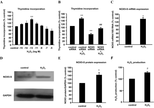
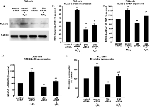
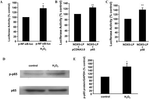
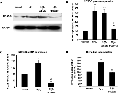
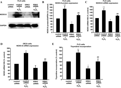
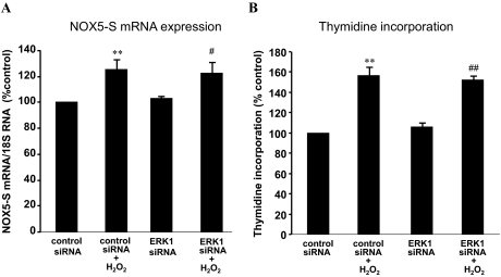
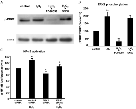
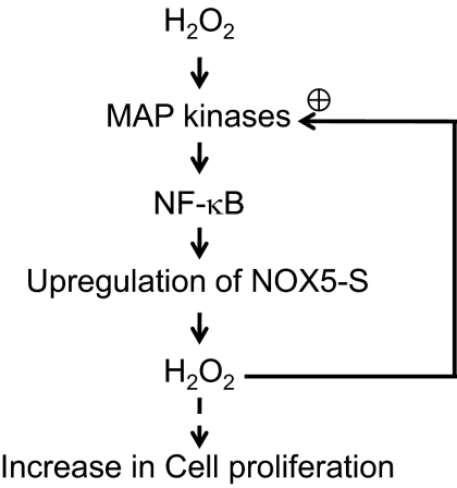
Mechanisms whereby acid reflux may accelerate the progression from Barrett's esophagus (BE) to esophageal adenocarcinoma (EA) are not fully understood. We have previously shown that NADPH oxidase NOX5-S generates reactive oxygen species (ROS) when Barrett's metaplastic cells are exposed to acid. Besides metaplastic cells, other H(2)O(2)-producing cells (e.g., inflammatory cells) present in BE mucosa may produce additional ROS, which may also affect metaplastic cells contributing to esophageal tumorigenesis. In this study, we investigate whether exogenous H(2)O(2) stimulates cell proliferation by increasing NOX5-S expression. Low dose (10(-13) M) of H(2)O(2) significantly increased thymidine incorporation, NOX5-S mRNA, and protein expression in a Barrett's EA cell line FLO. H(2)O(2)-induced increase in NOX5-S expression was significantly inhibited by knockdown of nuclear factor (NF)-κB1 p50 with p50 small interfering RNA (siRNA) in EA cell lines FLO and OE33. H(2)O(2) significantly increased p65 phosphorylation and the luciferase activity in FLO cells transfected with a NF-κB activation reporter plasmid pNF-κB-Luc. H(2)O(2)-induced increase in luciferase activity in FLO cells was significantly decreased by knockdown of extracellular signal-regulated kinase 2 (ERK2) mitogen-activated protein kinase (MAPK). Overexpression of p50 and p65 remarkably increased the luciferase activity in FLO cells transfected with a NOX5-S reporter plasmid NOX5-LP. In addition, H(2)O(2)-induced thymidine incorporation in FLO cells was significantly decreased by the MAPK kinase 1/2 inhibitor 2'-amino-3'methoxyflavone (PD98059) and ERK2 siRNA but not by ERK1 siRNA. Likewise, H(2)O(2)-induced increase in NOX5-S expression was significantly decreased by ERK2 siRNA in FLO and OE33 cells. We conclude that a low dose of H(2)O(2) increases cell proliferation. H(2)O(2)-induced increase in cell proliferation may depend on sequential activation of ERK2 MAPK, NF-κB1 p50, and NOX5-S.


Figures

Fig. 1.
Fig. 1.
H2O2 up-regulates NOX5-S expression in FLO cells. A, FLO cells were incubated with different concentrations of H2O2 (10−5, 10−7, 10−9, 10−11, 10−13, 10−14, 10−15 M) for 48 h and then incubated with methyl-[3H]thymidine (0.05 μCi/ml) for 4 h. H2O2 (10−13 M) significantly increased thymidine incorporation, whereas higher doses (10−5 and 10−7 M) slightly decreased thymidine incorporation. B, FLO cells were treated with H2O2 (10−13 M, 48 h) 24 h after NOX5 siRNA and control siRNA were introduced into FLO cells by Lipofectamine 2000. Knockdown of NOX5-S significantly decreased thymidine incorporation at basal condition and in response to H2O2 treatment. C, FLO cells were treated with 10−13 M H2O2 for 48 h, and then NOX5-S mRNA levels were measured by real-time PCR. H2O2 (10−13 M) significantly increased NOX5-S mRNA levels. D and E, a typical image of three Western blot analyses (D) and summarized data (E) show that 10−13 M H2O2 significantly increased NOX5-S protein level. FLO cells were treated with 10−13 M H2O2 for 48 h. F, FLO cells were treated with 10−13 M H2O2 for 48 h, washed, and cultured for an additional 24 h. H2O2 levels in culture medium were measured by using an Amplex Red H2O2 assay kit. A 48-h H2O2 treatment significantly increased H2O2 production (n = 3). t test, *, P < 0.05, #, P < 0.01; ANOVA, **, P < 0.01, compared with control or control siRNA group; ANOVA, ##, P < 0.01, compared with control siRNA plus H2O2 group.
Fig. 2.
Fig. 2.
Role of NF-κB in H2O2-induced NOX5-S expression. Transfection of p50 siRNA and control siRNA was carried out with Lipofectamine 2000. After a 4-h transfection, the transfection medium was replaced with regular medium. Twenty-four hours later, the transfectants were exposed to H2O2 (10−13 M) in fresh medium for an additional 48 h. A and B, a typical image of three Western blot analyses (A) and summarized data (B) show that knockdown of p50 significantly decreased NOX5-S protein expression at basal condition and in response to H2O2 treatment in FLO cells. C, knockdown of p50 significantly decreased NOX5 mRNA levels in response to H2O2 treatment in FLO cells. D, knockdown of p50 significantly decreased NOX5 mRNA levels in response to H2O2 treatment in OE33 cells. E, knockdown of p50 significantly decreased not only thymidine incorporation at basal condition but also H2O2-induced increase in thymidine incorporation in FLO cells. The data suggest that p50 mediates H2O2-induced increase in NOX5-S expression and in cell proliferation. n = 3, ANOVA, *, P < 0.05, **, P < 0.01, compared with control siRNA group; ANOVA. #, P < 0.05, compared with control siRNA + H2O2 group; ##, P < 0.01, compared with control siRNA group plus H2O2 group.
Fig. 3.
Fig. 3.
NF-κB1 p50 may be involved in H2O2-induced NOX5-S. A, twenty-four hours after transfection with pNF-κB-Luc plasmid and R. reniformis luciferase plasmid, FLO cells were treated with 10−13 M H2O2 for 48 h, and then luciferase activity was measured. H2O2 (10−13 M) significantly increased the luciferase activity in FLO cells, suggesting that H2O2 may activate NF-κB. B, twenty-four hours after transfection with NOX5-LP and R. reniformis luciferase plasmid plus p65 or pCDNA3.0 (control), luciferase activity was measured. Transfection with p65 expression plasmid significantly increased luciferase activity, suggesting that p65 may activate NOX5-S promoter. C, twenty-four hours after transfection with NOX5-LP and R. reniformis luciferase plasmid plus p50 or pCMV4 (control), luciferase activity was measured. Transfection with p50 expression plasmid significantly increased luciferase activity, suggesting that p50 may activate NOX5-S promoter. D and E, a typical image of three Western blot analyses (D) and summarized data (E) show that 10−13 M H2O2 significantly increased p65 phosphorylation. FLO cells were treated with 10−13 M H2O2 for 48 h. n = 3, t test, *, P < 0.05, compared with pNF-κB-Luc group or NOX5-LP plus pCDNA3.0 group; **, P < 0.01, compared with NOX5-LP or NOX5-LP + pCDNA3.0.
Fig. 4.
Fig. 4.
Role of MAPK in H2O2-induced NOX5-S expression. A and B, a typical image of three Western blot analyses (A) and summarized data (B) show that H2O2 significantly increased NOX5-S protein expression in FLO cells, an increase that was significantly decreased by PD98059. C, H2O2 significantly increased NOX5-S mRNA levels in FLO cells, an increase that was significantly decreased by PD98059, suggesting that MAPK may be involved in H2O2-induced NOX5-S expression. D, H2O2 significantly increased thymidine incorporation in FLO cells, an increase that was significantly decreased by PD98059, suggesting that MAPK may be involved in H2O2-induced increase in cell proliferation. n = 3, ANOVA, *, P < 0.05, **, P < 0.01, compared with control group; #, P < 0.05, compared with H2O2 + vehicle group; ##, P < 0.01, compared with H2O2 group.
Fig. 5.
Fig. 5.
ERK2 MAPK may be involved in H2O2-induced NOX5-S expression. Transfection of ERK2 siRNA and control siRNA was carried out with Lipofectamine 2000. After a 4-h transfection, the transfection medium was replaced with regular medium. Twenty-four hours later, the transfectants were exposed to H2O2 (10−3 M) in fresh medium for an additional 48 h. A and B, a typical image of three Western blot analyses (A) and summarized data (B) show that knockdown of ERK2 significantly decreased NOX5-S protein expression at basal condition and in response to H2O2 treatment in FLO cells. C, knockdown of ERK2 significantly decreased NOX5 mRNA levels at basal condition and in response to H2O2 treatment in FLO cells. D, knockdown of ERK2 significantly decreased NOX5 mRNA levels in response to H2O2 treatment in OE33 cells. E, knockdown of ERK2 significantly decreased thymidine incorporation at basal condition and in response to H2O2 treatment in FLO cells. The data suggest that ERK2 may mediate H2O2-induced increase in NOX5-S expression and in cell proliferation. n = 3, ANOVA, *, P < 0.05, **, P < 0.01, compared with control siRNA group; #, P < 0.05, ##, P < 0.01, compared with control siRNA plus H2O2 group.
Fig. 6.
Fig. 6.
ERK1 MAPK may be not involved in H2O2-induced NOX5-S expression. Transfection of ERK1 siRNA and control siRNA was carried out with Lipofectamine 2000. After a 4-h transfection, the transfection medium was replaced with regular medium. Twenty-four hours later, the transfectants were exposed to H2O2 (10−13 M) in fresh medium for an additional 48 h. A, knockdown of ERK1 had no effect on H2O2-induced NOX5 mRNA levels. B, knockdown of ERK1 had no effect on H2O2-induced increase in thymidine incorporation. The data suggest that ERK1 may not be involved in H2O2-induced increase in NOX5-S expression and in cell proliferation. n = 3, ANOVA, **, P < 0.01, compared with control siRNA group; #, P < 0.05, ##, P < 0.01, compared with control siRNA plus H2O2 group.
Fig. 7.
Fig. 7.
H2O2 may activate ERK2 MAPK. A and B, a typical image of three Western blot analyses (A) and summarized data (B) show that H2O2 significantly increased ERK2 phosphorylation, an increase that was significantly decreased by PD98059 (10−5 M) but not by SN50 (10−5 M). FLO cells were exposed to 10−5 M H2O2 for 15 min in the absence or presence of PD98059 (10−5 M) or SN50 (10−5 M). C, FLO cells were transfected with 0.1 μg of R. reniformis luciferase plasmid and 0.5 μg of pNF-κB-Luc plasmid by using Lipofectamine 2000. Twenty-four hours after transfection with plasmids, 60 pmol of ERK2 or control siRNA per well were transfected. Twenty-four hours later, FLO cells were treated with 10−13 M H2O2 for additional 48 h. Finally, the luciferase activity was measured. H2O2 significantly increased luciferase activity in FLO cells, an increase that was significantly reduced by knockdown of ERK2, suggesting that activation of NF-κB may depend on activation of ERK2. n = 3. **, P < 0.01, ANOVA, compared with control siRNA group; *, P < 0.05, ANOVA, compared with control siRNA group; #, P < 0.05, ANOVA, compared with control siRNA plus H2O2 group; ##, P < 0.01, ANOVA, compared with H2O2 group.
Fig. 8.
Fig. 8.
Signaling in H2O2-induced increase in cell proliferation. Low doses of H2O2 increase cell proliferation. H2O2-induced increase in cell proliferation may depend on sequential activation of ERK2 MAPK and NF-κB and thus up-regulate NOX5-S expression, which further enhances production of ROS (a positive feedback).

Similar articles

Cited by

References

    1. Ballard DW, Dixon EP, Peffer NJ, Bogerd H, Doerre S, Stein B, Greene WC. (1992) The 65-kDa subunit of human NF-kappa B functions as a potent transcriptional activator and a target for v-Rel-mediated repression. Proc Natl Acad Sci USA 89:1875–1879 - PMC - PubMed
    1. Bánfi B, Maturana A, Jaconi S, Arnaudeau S, Laforge T, Sinha B, Ligeti E, Demaurex N, Krause KH. (2000) A mammalian H+ channel generated through alternative splicing of the NADPH oxidase homolog NOH-1. Science 287:138–142 - PubMed
    1. Blot WJ, McLaughlin JK. (1999) The changing epidemiology of esophageal cancer. Semin Oncol 26 (5 Suppl 15):2–8 - PubMed
    1. Bradford MM. (1976) A rapid and sensitive method for the quantitation of microgram quantities of protein utilizing the principle of protein-dye binding. Anal Biochem 72:248–254 - PubMed
    1. Cao W, Sohn UD, Bitar KN, Behar J, Biancani P, Harnett KM. (2003) MAPK mediates PKC-dependent contraction of cat esophageal and lower esophageal sphincter circular smooth muscle. Am J Physiol Gastrointest Liver Physiol 285:G86–G95 - PubMed

Publication types

MeSH terms