. 2011 Oct;90(4):787-97.
doi: 10.1189/jlb.0111014. Epub 2011 Jul 12.

Role of TLR signaling in Francisella tularensis-LPS-induced, antibody-mediated protection against Francisella tularensis challenge

Affiliations

Role of TLR signaling in Francisella tularensis-LPS-induced, antibody-mediated protection against Francisella tularensis challenge

Leah E Cole et al. J Leukoc Biol. 2011 Oct.

Abstract

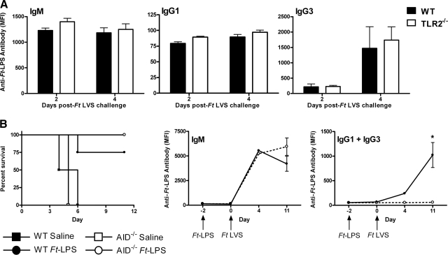
Immunization with Ft-LPS provokes an antigen-specific, B-1a cell-derived antibody response that protects WT mice against an otherwise lethal challenge with Ft LVS. However, this same regimen offers limited protection to TLR2(-/-) mice, despite production of WT levels of anti-Ft-LPS antibodies. As Ft-LPS exhibits no TLR2 agonist activity, and macrophage-induced cytokine production in response to Ft LVS is overwhelmingly TLR2-dependent, we hypothesized that treatment of TLR2(-/-) mice with an alternative, MyD88-dependent TLR agonist would compensate for reduced recognition of Ft LVS in TLR2(-/-) mice and thereby, restore Ft-LPS-mediated protection. Administration of the nontoxic TLR4 agonist, synthetic Escherichia coli MPL, at the time of Ft-LPS immunization or Ft LVS challenge, fully protected TLR2(-/-) mice, whereas treatment of WT or TLR2(-/-) mice with MPL alone conferred partial protection. The TLR5 agonist, flagellin, also synergized with Ft-LPS to protect TLR2(-/-) mice from lethal Ft LVS challenge. In contrast to Ft LVS, Ft-LPS pretreatment failed to protect mice against i.n. challenge with Ft Schu S4, whereas MPL, administered in the absence or presence of Ft-LPS, conferred significant, albeit partial, protection. MPL treatment of macrophages increased the uptake of Ft LVS and decreased intracellular bacterial survival while shifting the macrophage-differentiation phenotype from "alternatively activated" to "classically activated". Collectively, our data suggest that optimal, Ft-LPS-mediated protection against Ft LVS infection requires two discrete events, i.e., production of Ft-LPS-specific antibody, as well as TLR-mediated macrophage activation, to fully control Francisella infection.


Figures

Figure 1.
Figure 1.. Pretreatment with Ft-LPS provides minimal protection in TLR2−/− and IRAK4KD-KI mice against subsequent Ft LVS challenge.
Female WT (C57BL/6J), TLR2−/− (A), or IRAK4KD-KI (B) mice were pretreated with saline or Ft-LPS (100 ng), 2 days prior to i.p. challenge with Ft LVS, as described in Materials and Methods. The survival curves for the Ft-LPS-pretreated animals (WT vs. TLR2−/−, log-rank test, P<0.0001; and WT vs. IRAK4KD-KI, log-rank test, P=0.0004) are significantly different. Graphs represent the combined results of four or three separate experiments for A and B, respectively.
Figure 2.
Figure 2.. The failure of TLR2−/− mice to be protected fully by Ft-LPS is not attributable to a failure to produce anti-LPS antibodies and requires IgM only.
(A) TLR2−/− mice produce anti-Ft-LPS antibodies at levels comparable to that of WT mice. Groups of WT and TLR2−/− mice were injected i.p. with Ft-LPS (100 ng), 3 days prior to i.p. challenge with 9 × 10³ CFU Ft LVS. Two and 4 days after bacterial challenge, mice were bled and the sera analyzed for the presence of anti-Ft-LPS IgM, IgG1, and IgG3. Data presented are derived from a single experiment with five mice/treatment group, which is representative of five independent experiments with similar design and outcome. There were no significant differences between the response of WT and TLR2−/− mice at either time-point for each isotype analyzed. MFI, Mean fluorescence intensity. (B) Isotype-switching is not required for Ft-LPS-induced protection against Ft LVS challenge. WT or AID−/− mice were pretreated with saline or Ft-LPS (100 ng), 2 days prior to i.p. challenge with 6.5 × 10³ CFU Ft LVS, as described in A. (Left panel) Pretreatment with Ft-LPS protected WT and AID−/− mice against subsequent Ft LVS challenge. (Middle and right panels) Sera were collected from all animals on Day –2, prior to Ft-LPS immunization; on Day 0, prior to Ft LVS challenge; and from healthy survivors, 4 and 11 days post-challenge. Sera were analyzed for the presence of anti-Ft-LPS IgM (middle panel) and IgG1 and IgG3 (right panel). Data are derived from a single experiment that is representative of three independent experiments of similar design and outcome; *P = 0.007.
Figure 3.
Figure 3.. Treatment with MPL, a TLR4 agonist, prior to or at the time of Ft LVS challenge, restores full protective capacity of Ft-LPS pretreatment in TLR2−/− mice.
Groups of four or five WT (A) or TLR2−/− (B) mice were injected i.p. with saline or Ft-LPS (100 ng), 2 days prior to i.p. challenge with Ft LVS, as described in Materials and Methods. MPL (100 μg) was administered i.p., concurrently with Ft-LPS pretreatment (Day –2) or at the time of bacterial challenge (Day 0). These data represent the combined results from four independent experiments. For WT mice: saline versus Ft-LPS (P<0.0001); saline versus Ft-LPS, MPL (Day –2; P=0.0011); saline versus Ft-LPS, MPL (Day 0; P<0.0001); saline versus MPL (P<0.0001); and MPL versus Ft-LPS, MPL (P=0.048). For TLR2−/− mice: saline vs. Ft-LPS (P<0.0001); saline versus Ft-LPS, MPL (Day –2; P=0.001); saline versus Ft-LPS, MPL (Day 0; P<0.001); saline versus MPL (P=0.0045); and MPL versus Ft-LPS, MPL (Day 0; P=0.0275). (C) TLR2−/− mice were injected i.p. with saline, Ft-LPS, MPL (100 μg), and challenged with saline or Ft LVS at the times indicated on the graph. Two days after Ft LVS challenge, all mice were killed, and RNA was extracted from the livers. Levels of Ft LVS 16S rRNA mRNA were measured as described previously [16]. These data represent the mean ± sem (eight to 10 mice/treatment) derived from three independent experiments. *P < 0.001 versus saline (Day –2)/Ft LVS (Day 0); #P < 0.01 versus saline (Day –2)/Ft LVS (Day 0).
Figure 4.
Figure 4.. Treatment with flagellin, a TLR5 agonist, prior to Ft LVS challenge, restores full protective capacity of Ft-LPS pretreatment in TLR2−/− mice.
Groups of four TLR2−/− mice were injected i.p. with saline, Ft-LPS (100 ng), flagellin (5 μg), or Ft-LPS and flagellin, 2 days prior to i.p. challenge with Ft LVS (25,500 CFU), as described in Materials and Methods. The survival data (A) and survivor weight loss data (B) represent the mean ± sem of a single experiment that is representative of three independent experiments of similar design and outcome. For the survival study, saline versus flagellin (P=0.63); saline versus Ft-LPS (P<0.007); saline versus Ft-LPS + flagellin (P=0.007).
Figure 5.
Figure 5.. MPL treatment leads to minimal induction of anti-Ft-LPS antibody.
(A) Groups of five WT or TLR2−/− mice were injected i.p. with saline, MPL (100 μg), or Ft-LPS (100 ng). Mice were bled prior to treatment (Prebleed), as well as 5 and 12 days after initial injection. All sera were analyzed for the presence of anti-Ft-LPS IgM. Data presented are derived from a single experiment, representative of three independent experiments with similar design and outcome. (B) Groups of five WT or TLR2−/− mice were injected i.p. with saline, Ft-LPS (100 ng), or MPL (100 μg) at the indicated times and were challenged on Day 0 with Ft LVS. Two days later, mice were bled and the sera analyzed for the presence of anti-Ft-LPS IgM. Data are derived from a single experiment that is representative of three independent experiments of similar design and outcome. Data shown are the mean ± sem. No significant differences were observed at any time-point between WT and TLR2−/− mice.
Figure 6.
Figure 6.. MPL treatment favors induction of classically versus alternatively activated macrophages and results in enhanced intracellular killing of Ft LVS.
WT macrophage cultures were treated in triplicate for 1 h with MPL (100 ng/ml), followed by infection with Ft-LPS (MOI=40), gentamicin treatment to kill extracellular bacteria, and then incubation in medium only for an additional 24 h. Total cellular RNA was subjected to qPCR, as described in Materials and Methods, for the detection of iNOS and arginase-1 mRNA (A) and for the detection of Ft 16S rRNA (B). Results represent the combined data from two independent experiments; *P < 0.05. (C) MPL enhances expression of IFN-β mRNA in uninfected and Ft LVS-infected macrophages. The experimental approach is identical to that described in A and B, with additional time-points as indicated in the figure. P < 0.05 for medium versus MPL at each time-point examined.

Similar articles

Cited by

References

    1. Saslaw S., Eigelsbach H. T., Wilson H. E., Prior J. A., Carhart S. (1961) Tularemia vaccine study. I. Intracutaneous challenge. Arch. Intern. Med. 107, 689–701 - PubMed
    1. Saslaw S., Eigelsbach H. T., Prior J. A., Wilson H. E., Carhart S. (1961) Tularemia vaccine study. II. Respiratory challenge. Arch. Intern. Med. 107, 702–714 - PubMed
    1. Christopher G. W., Cieslak T. J., Pavlin J. A., Eitzen E. M., Jr. (1997) Biological warfare. A historical perspective. JAMA 278, 412–417 - PubMed
    1. Dennis D. T., Inglesby T. V., Henderson D. A., Bartlett J. G., Ascher M. S., Eitzen E., Fine A. D., Friedlander A. M., Hauer J., Layton M., Lillibridge S. R., McDade J. E., Osterholm M. T., O′Toole T., Parker G., Perl T. M., Russell P. K., Tonat K. (2001) Tularemia as a biological weapon: medical and public health management. JAMA 285, 2763–2773 - PubMed
    1. Harris S. (1992) Japanese biological warfare research on humans: a case study of microbiology and ethics. Ann. N. Y. Acad. Sci. 666, 21–52 - PubMed

Publication types

MeSH terms