

Magnesium reduces calcification in bovine vascular smooth muscle cells in a dose-dependent manner
- PMID: 21750166
- PMCID: PMC3275783
- DOI: 10.1093/ndt/gfr321
Magnesium reduces calcification in bovine vascular smooth muscle cells in a dose-dependent manner
Abstract
Background: Vascular calcification (VC), mainly due to elevated phosphate levels, is one major problem in patients suffering from chronic kidney disease. In clinical studies, an inverse relationship between serum magnesium and VC has been reported. However, there is only few information about the influence of magnesium on calcification on a cellular level available. Therefore, we investigated the effect of magnesium on calcification induced by β-glycerophosphate (BGP) in bovine vascular smooth muscle cells (BVSMCs).
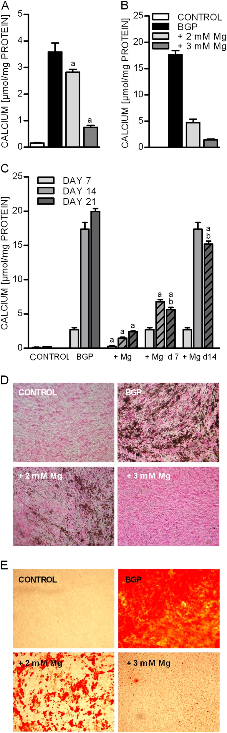
Methods: BVSMCs were incubated with calcification media for 14 days while simultaneously increasing the magnesium concentration. Calcium deposition, transdifferentiation of cells and apoptosis were measured applying quantification of calcium, von Kossa and Alizarin red staining, real-time reverse transcription-polymerase chain reaction and annexin V staining, respectively.
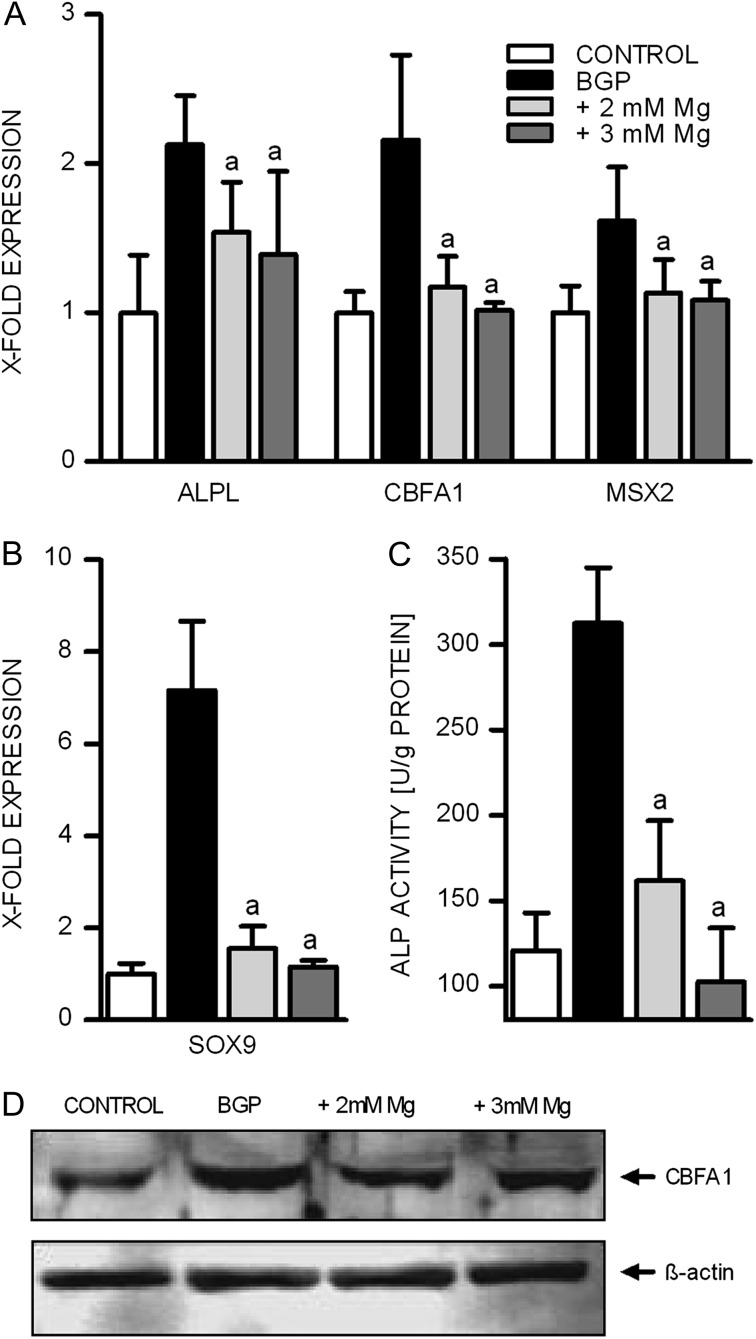
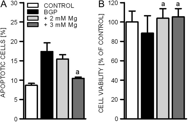
Results: Calcium deposition in the cells dramatically increased with addition of BGP and could be mostly prevented by co-incubation with magnesium. Higher magnesium levels led to inhibition of BGP-induced alkaline phosphatase activity as well as to a decreased expression of genes associated with the process of transdifferentiation of BVSMCs into osteoblast-like cells. Furthermore, estimated calcium entry into the cells decreased with increasing magnesium concentrations in the media. In addition, higher magnesium concentrations prevented cell damage (apoptosis) induced by BGP as well as progression of already established calcification.
Conclusions: Higher magnesium levels prevented BVSMC calcification, inhibited expression of osteogenic proteins, apoptosis and further progression of already established calcification. Thus, magnesium is influencing molecular processes associated with VC and may have the potential to play a role for VC also in clinical situations.
Figures

References
-
- Blacher J, Guerin AP, Pannier B, et al. Arterial calcifications, arterial stiffness, and cardiovascular risk in end-stage renal disease. Hypertension. 2001;38:938–942. - PubMed
-
- Goodman WG, Goldin J, Kuizon BD, et al. Coronary-artery calcification in young adults with end-stage renal disease who are undergoing dialysis. N Engl J Med. 2000;342:1478–1483. - PubMed
-
- Covic A, Kanbay M, Voroneanu L, et al. Vascular calcification in chronic kidney disease. Clin Sci (Lond) 2010;119:111–121. - PubMed
-
- Moe SM, Chen NX. Pathophysiology of vascular calcification in chronic kidney disease. Circ Res. 2004;95:560–567. - PubMed
-
- Ishimura E, Okuno S, Kitatani K, et al. Significant association between the presence of peripheral vascular calcification and lower serum magnesium in hemodialysis patients. Clin Nephrol. 2007;68:222–227. - PubMed
Publication types
MeSH terms
Substances
LinkOut - more resources
Full Text Sources
Other Literature Sources
Miscellaneous
