. 2011 Aug 1;124(Pt 15):2666-75.
doi: 10.1242/jcs.085480. Epub 2011 Jul 12.

MMP9 cleavage of the β4 integrin ectodomain leads to recurrent epithelial erosions in mice

Affiliations

MMP9 cleavage of the β4 integrin ectodomain leads to recurrent epithelial erosions in mice

Sonali Pal-Ghosh et al. J Cell Sci. .

Abstract

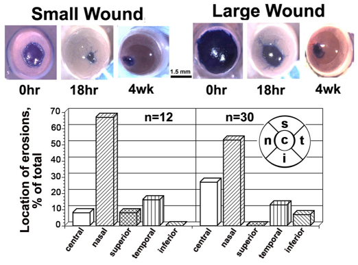
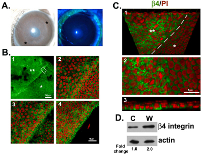
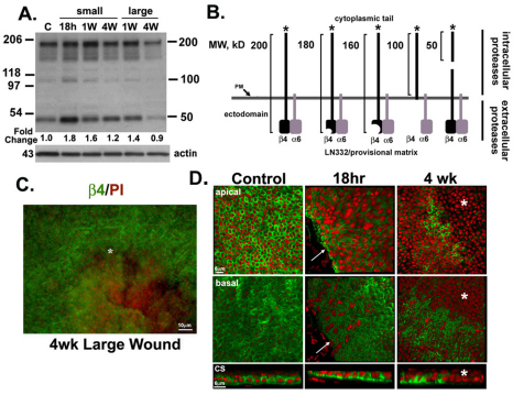
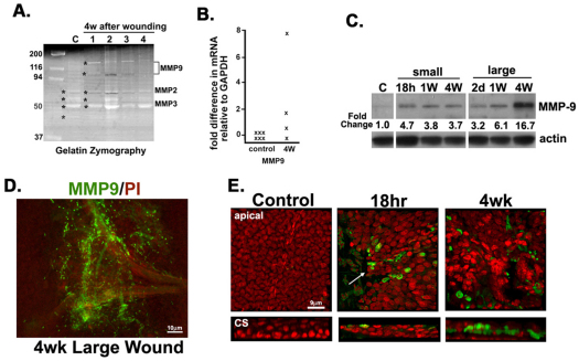
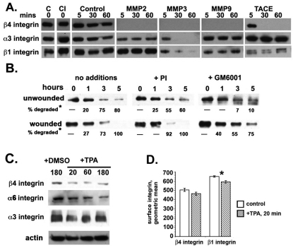
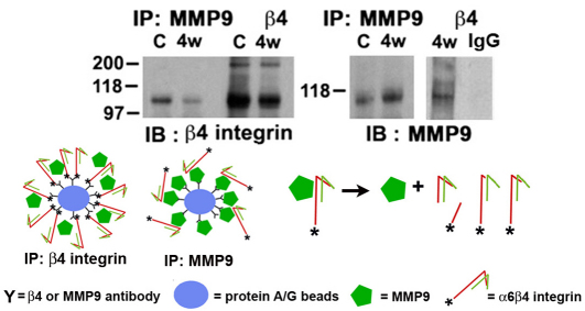
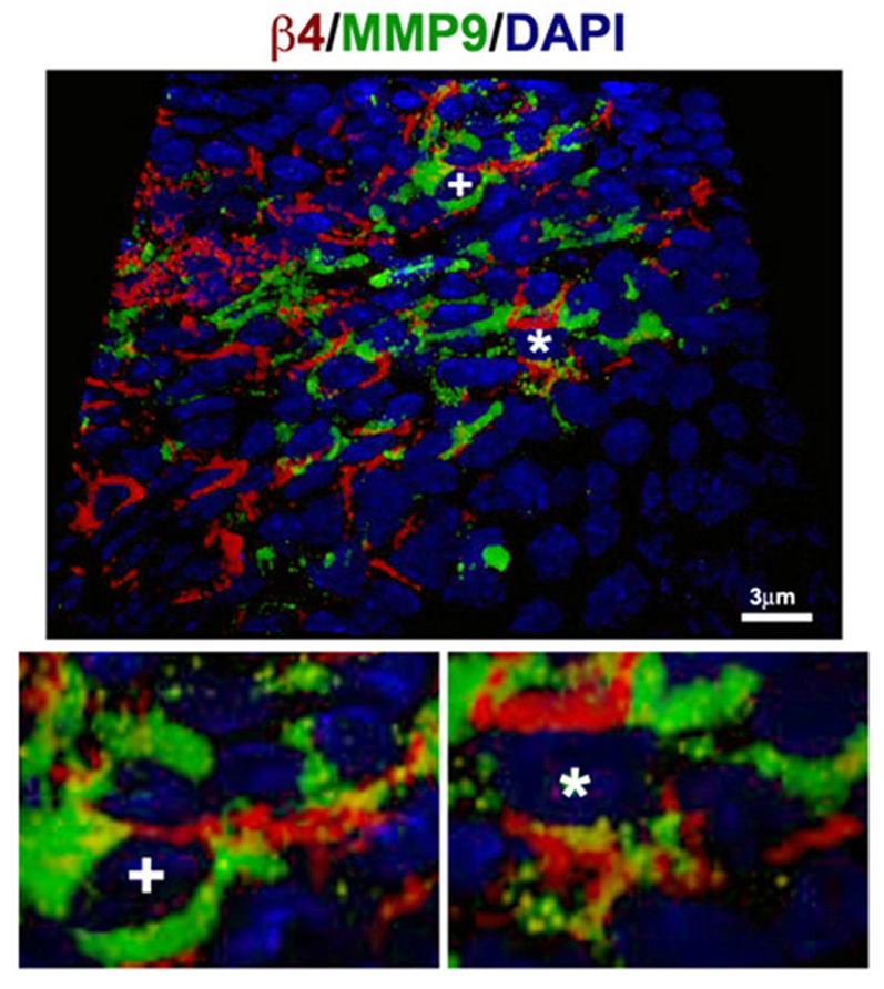
Integrin α6β4 is an integral membrane protein within hemidesmosomes and it mediates adhesion of epithelial cells to their underlying basement membrane. During wound healing, disassembly of hemidesmosomes must occur for sheet movement-mediated cell migration. The mechanisms of disassembly and reassembly of hemidesmosomes are not fully understood. The current study was initiated to understand the underlying cause of recurrent corneal erosions in the mouse. Here, we show that in vivo: (1) MMP9 levels are elevated and β4 integrin is partially cleaved in epithelial cell extracts derived from debridement wounded corneas; (2) the β4 ectodomain is missing from sites where erosions develop; and (3) β4 cleavage can be reduced by inhibiting MMP activity. Although β4, α3 and β1 integrins were all cleaved by several MMPs, only MMP9 was elevated in cell extracts derived from corneas with erosions. Coimmunoprecipitation studies showed that β4 integrin associates with MMP9, and protein clustering during immunoprecipitation induced proteolytic cleavage of the β4 integrin extracellular domain, generating a 100 kDa β4 integrin cytoplasmic domain fragment. Confocal imaging with three-dimensional reconstruction showed that MMP9 localizes at erosion sites in vivo where the ectodomain of β4 integrin is reduced or absent. MMP activation experiments using cultured corneal and epidermal keratinocytes showed reduced levels of α6β4 and β1 integrins within 20 minutes of phorbol ester treatment. This report is the first to show that β4 integrin associates with MMP9 and that its ectodomain is a target for cleavage by MMP9 in vivo under pathological conditions.


Figures

Fig. 1.
Fig. 1.
Epithelial sheets move rapidly to close debridement wounds, but over time, recurrent erosions develop. Open wounds detected by Richardson stain at various times after wounding are shown in the top panel. Scale bar: 1.5 mm. The anatomical locations of the erosions that develop are shown graphically below as the percentage of the erosions that develop centrally or at the nasal, superior, temporal or inferior quadrants. Using Chi-squared statistics, after both small and large wounds, there are significantly more erosions in the periphery compared with the center of the cornea. In the periphery, there are more erosions in the nasal quadrant compared with all other quadrants combined (P<0.001).
Fig. 2.
Fig. 2.
Basement membrane removal prevents erosions and enhances β4 integrin localization and expression within the injury site. (A) Keratectomy wounded eyes 4 weeks after wounding. Eyes were evaluated for open wounds with both slit lamp and 2% fluorescein eye drops. The two asterisks in the slit lamp image indicate the margins of the wound seen as a scar after 4 weeks. (B) β4 staining of 4-week cornea in green at 10× magnification (1). The double white asterisks indicate the keratectomy-wounded area and single asterisk, the unwounded area. (2) A 63× image of boxed area in 1. Images 2–4 are taken from apical to basal at 2–3 μm intervals. (C) 3-D reconstruction of a keratectomy wounded area at 63× (1). The image has been tilted at a 45° angle so the β4 staining can be seen basally. The dotted lines indicate the boundary between wounded and unwounded area. Image 2 is a magnified view of 1, showing the increased expression of β4 and image 3 is a cross-sectional view of 1 highlighting the increased number of cell layers at the keratectomy site. (D) Immunoblots of corneal epithelium from control and keratectomy corneas show that 4-week keratectomy-wounded corneal epithelia express more β4 integrin than control epithelia.
Fig. 3.
Fig. 3.
The appearance of β4 integrin degradation products during healing of debridement wounds is accompanied by the loss of the β4 integrin ectodomain adjacent to erosions. (A) Immunoblots of corneal epithelia harvested at different time points probed with β4 integrin and actin showing the fold change in β4 integrin expression over time compared with control. Blots show formation of proteolytic cleavage products derived from β4 integrin during wound healing of small and large wounds. Quantification of these bands are shown in supplementary material Fig. S1. (B) Schematic representation of the types of cleavage products expected from cleavage of β4 integrin into different size fragments. Fragments over 100 kDa, are derived from the action of extracellular proteases whereas fragment less than 100 kDa are derived from intracellular proteases. (C)10× image showing the loss of β4 at erosion site (*) 4 weeks after wounding with β4 in green and PI in red. (D) 3D reconstruction of 63× images of control, 18 hour and 4 week tissues stained with β4 and PI showing apical, basal and cross sectional (CS) views of the corneal epithelium. The white arrows indicate the leading edge at 18 hours and the asterisks indicate the erosion site with loss of β4 integrin.
Fig. 4.
Fig. 4.
MMP9 is upregulated at the mRNA and protein levels in corneas with erosions. (A) Gelatin zymography of four epithelial tissue extracts taken 4 weeks after wounding compared with a control extract shows increased MMP activity for proteins with molecular mass consistent with that of MMP9. Asterisks indicate the mobility of each of the major bands detected. (B) QPCR performed on mRNA isolated from corneal epithelia shows below detectable mRNA levels for MMP9 before wounding in all control samples evaluated (n=6 animals) and increased levels in three of four samples after wounding. Each ‘×’ represents RNA from two corneas and data are expressed relative to Gapdh mRNA levels. (C) Immunoblots of corneal epithelial samples at different time points probed with MMP9 and actin showing the fold change in MMP9 expression compared with control. (D)10× magnification image of the erosion site indicating increased MMP9 in green. (E) 3D reconstruction of 63× images at control, 18 hours and 4 weeks after wounding shown en face and in cross section (CS). The arrow in the 18 hour image indicates the MMP9-positive cells at the leading edge. The area adjacent to the erosion site at 4 weeks (4W) shows a loss of tissue organization and increased levels of MMP9 expression.
Fig. 5.
Fig. 5.
MMPs and TACE cleave β4 integrin, which can be partially inhibited by GM6001 and protease inhibitors, and TPA induces a rapid loss of α6 and β4, but not α3 integrin. (A) The study of MMP-specific degradation of integrins by MMPs and TACE at 5, 30 and 60 minutes. A longer exposure of the β4 integrin data is provided in supplementary material Fig. S2. (B) Extracts from unwounded and 18-hour-wounded corneas were used to compare the rate of cleavage of β4 integrin with and without addition of a protease inhibitor cocktail or the MMP inhibitor GM6001. The asterisk indicates that data were quantified and the percentage degradation calculated relative to the amount of β4 integrin present at time 0. For unwounded GM6001-treated extracts, both β4 bands were summed for the calculations. Data show that in unwounded corneas, GM6001 can block most of the β4 integrin degradation, but can only delay degradation in wounded corneas. (C) MMP activation was induced by TPA and integrin degradation studied in human corneal epithelial cells. Cells were treated with 0.02% DMSO for 180 minutes as controls and with 0.02% DMSO plus TPA for 20, 60 and 180 minutes. Data show a rapid decrease in all three integrins studied at 20 minutes; by 180 minutes, α3 integrin expression partially recovered, whereas expression of β4 and α6 integrin remained lower than DMSO-treated control cells. (D) MMP activation was induced by TPA and β4 and β1 integrin ectodomain shedding studied in primary mouse epidermal keratinocytes 20 minutes after treatment using flow cytometry. Data indicate a significant reduction in β1 integrin on cell surfaces after MMP activation (*P<0.05); the reduction in β4 surface expression was not quite statistically significant.
Fig. 6.
Fig. 6.
MMP9 coimmunoprecipitates with and cleaves full-length β4 integrin. Control (C) and wounded (4w) tissues were immunoprecipitated with antibodies against the β4 integrin cytoplasmic domain and MMP9. Blots probed with the β4 cytoplasmic domain antibody reveal that MMP9 and full-length β4 integrin associate with one another and this association induces β4 integrin cleavage. Blots probed with antibody against MMP9 reveal that MMP9 is present in β4 integrin immunoprecipitates. Beads incubated with control IgG are also shown. The diagram in the lower panel schematically shows protein A/G beads coated with either β4 integrin or MMP9 antibodies and incubated with corneal epithelial cell extracts containing α6β4 and MMP9. MMP9 associates with the β4 integrin extracellular domain and cleaves it. The expected degradation products are shown on the left. The symbols used to represent protein A/G, MMP9 and β4 integrin are also shown.
Fig. 7.
Fig. 7.
MMP9 induces β4 integrin ectodomain cleavage in vivo at sites of erosions. 3D image at 63× magnification of a 4-week cornea with erosion. The areas labeled with ‘+’ and ‘*’ have been digitally magnified 3× to reveal times focal sites of colocalization of MMP9 and β4 integrin ectodomain.

Similar articles

Cited by

References

    1. Borradori L., Sonnenberg A. (1996). Hemidesmosomes: roles in adhesion, signaling and human diseases. Curr. Opin. Cell Biol. 8, 647-656 - PubMed
    1. Chen P., Abacherli L. E., Nadler S. T., Wang Y., Li Q., Parks W. C. (2009). MMP7 shedding of syndecan-1 facilitates re-epithelialization by affecting α2β1 integrin activation. PLoS ONE 4, e6565 - PMC - PubMed
    1. Chetty C., Lakka S. S., Bhoopathi P., Rao J. S. (2010). MMP-2 alters VEGF expression via αvβ3 integrin-mediated PI3K/AKT signaling in A549 lung cancer cells. Int. J. Cancer 127, 1081-1095 - PMC - PubMed
    1. Choi W. S., Jeon O. H., Kim D. S. (2010). CD40 ligand shedding is regulated by interaction between matrix metalloproteinase-2 and platelet integrin αIIbβ3. J. Thromb. Haemost. 8, 1364-1371 - PubMed
    1. Chotikavanich S., de Paiva C. S., Li de-Q., Chen J. J., Bian F., Farley W. J., Pflugfelder S. C. (2009). Production and activity of matrix metalloproteinase-9 on the ocular surface increase in dysfunctional tear syndrome. Invest. Ophthalmol. Vis. Sci. 50, 3203-3209 - PMC - PubMed

Publication types