. 2011 Sep;10(9):1686-97.
doi: 10.1158/1535-7163.MCT-10-1108. Epub 2011 Jul 12.

Carfilzomib interacts synergistically with histone deacetylase inhibitors in mantle cell lymphoma cells in vitro and in vivo

Affiliations

Carfilzomib interacts synergistically with histone deacetylase inhibitors in mantle cell lymphoma cells in vitro and in vivo

Girija Dasmahapatra et al. Mol Cancer Ther. 2011 Sep.

Retraction in

Abstract

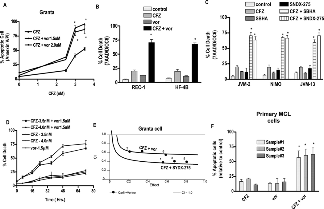
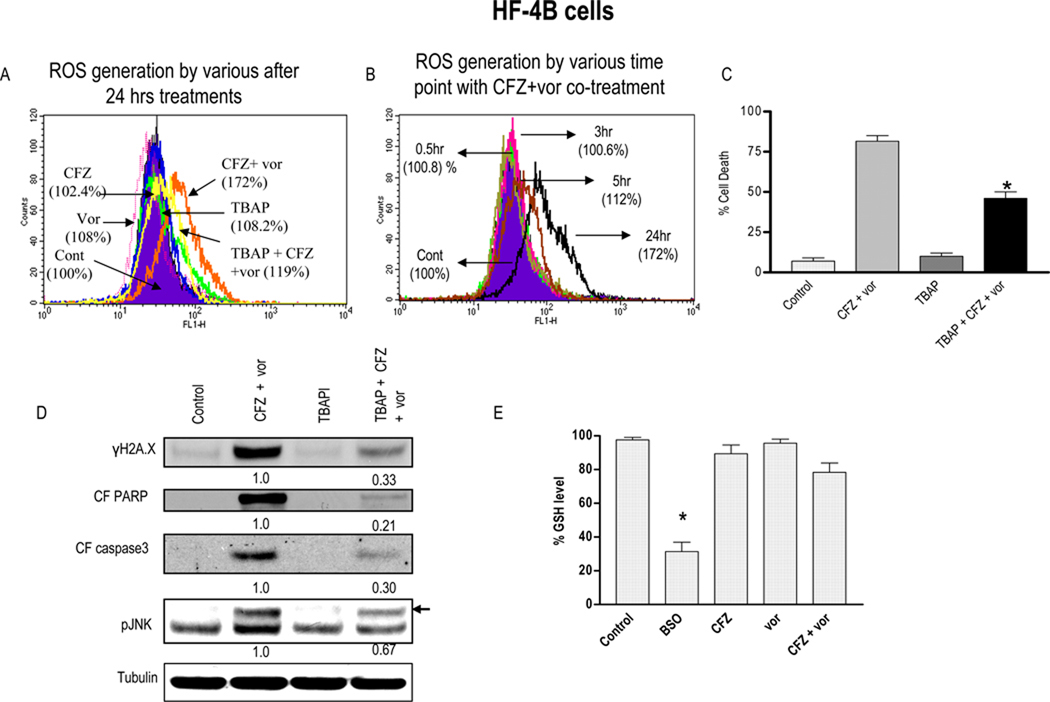
Interactions between the proteasome inhibitor carfilzomib and the histone deacetylase (HDAC) inhibitors vorinostat and SNDX-275 were examined in mantle cell lymphoma (MCL) cells in vitro and in vivo. Coadministration of very low, marginally toxic carfilzomib concentrations (e.g., 3-4 nmol/L) with minimally lethal vorinostat or SNDX-275 concentrations induced sharp increases in mitochondrial injury and apoptosis in multiple MCL cell lines and primary MCL cells. Enhanced lethality was associated with c-jun-NH,-kinase (JNK) 1/2 activation, increased DNA damage (induction of λH2A.X), and ERK1/2 and AKT1/2 inactivation. Coadministration of carfilzomib and histone deacetylase inhibitors (HDACI) induced a marked increase in reactive oxygen species (ROS) generation and G(2)-M arrest. Significantly, the free radical scavenger tetrakis(4-benzoic acid) porphyrin (TBAP) blocked carfilzomib/HDACI-mediated ROS generation, λH2A.X formation, JNK1/2 activation, and lethality. Genetic (short hairpin RNA) knockdown of JNK1/2 significantly attenuated carfilzomib/HDACI-induced apoptosis, but did not prevent ROS generation or DNA damage. Carfilzomib/HDACI regimens were also active against bortezomib-resistant MCL cells. Finally, carfilzomib/vorinostat coadministration resulted in a pronounced reduction in tumor growth compared with single agent treatment in an MCL xenograft model associated with enhanced apoptosis, λH2A.X formation, and JNK activation. Collectively, these findings suggest that carfilzomib/HDACI regimens warrant attention in MCL.


Conflict of interest statement

Potential conflict of interest: No conflict of interest to report

Figures

Figure 1
Figure 1. Co-treatment with carfilzomib and HDACIs leads to synergistic induction of cell death in various mantle cell lymphoma lines and primary mantle cell lymphoma cells
(A). Granta cells were treated with varying carfilzomib (CFZ) (1.0 – 4.0 nM) in with fixed vorinostat (vor) (1.5 or 0.2.0 µM) concentrations for 48 hr, after which apoptosis was monitored by Annexin V/PI staining. (B) REC-1 and HF-4B cells were treated with varying vorinostat (1.0–2.0 µM) concentrations ± fixed carfilzomib concentrations (2.5 or 3.0 nM respectively) for 36 h, after which cell death was monitored by 7AAD/DiOC6 staining (C) JVM-2, NIMO and JVM-13 cells treated with minimally toxic carfilzomib concentrations (JVM-2–4nM, NIMO-5nM, JVM-13-4nM) ± SBHA (JVM-2–30 µM, NIMO-50 µM, JVM-13–40 µM) and SNDX-275 (1.0 µM for JVM-2 and NIMO, 1.5µM for JVM-13) for 48 hours, after which cell death was monitored by 7-AAD and DiOC6 staining. (D) Granta cells were treated with the indicated concentration of carfilzomib and vorinostat and cell death at various intervals up to 72 hrs was monitored by 7AAD/DiOC6 staining. (E) Combination Index (C.I.) values were determined using CalcuSyn software as per program instruction (24). (F) Primary human MCL cells were isolated as described in Methods. They were treated with carfilzomib (1st sample - 1.5 nM, 2nd sample - 15 nM, 3rd sample - 5 nM) ± vorinostat (1st sample - 0.75µM, 2nd sample - 1.25 µM, 3rd sample - 0.5 µM) for 14 h. The percentage of apoptotic cells was monitored by Annexin V/PI staining and the percentage of dead cells was normalized to controls. Viability of the three primary specimens without treatment was 75–80%, 65–75%, 60–70% for three samples respectively. For all studies, values represent the means for 3 experiments performed in triplicate ± S.D. For A–C, F * = values significantly greater than those obtained with carfilzomib or vorinostat treatment alone; P < 0.01.
Figure 2
Figure 2. Combined treatment with carfilzomib or ONX0912 with vorinostat in HF-4B and Granta cells sharply increases caspase activation, PARP cleavage, JNK activation, MnSOD2 induction, and DNA damage
(A–B) HF-4B cells were treated with carfilzomib (4.0 nM) ± vorinostat (1.5µM) for 24hrs. (C) Granta cells were treated with carfilzomib (3.5 nM) ± vorinostat (1.5µM) for 24hrs (D) HF-4B cells were treated with ONX0912 (300 nM) ± vorinostat (1.5µM) for 24hrs. (E) HF-4B cells were pretreated with 10µM BOC-fmk for 30 minutes followed by carfilzomib (3.5 nM) ± vorinostat (1.5µM) for 24 hrs. Protein expression was monitored by Western blotting as described in Methods (24).
Figure 3
Figure 3. Co-administration of carfilzomib and vorinostat triggers ROS generation and pre-treatment with anti-oxidant TBAP partially blocks carfilzomib/vorinostat-mediated ROS generation and lethality
(A) Granta cells were treated with carfilzomib-3.5 nM ± vorinostat-1.5µM (± pre-treatment with 400 µM TBAP for 3hrs) for 24 hrs after which ROS generation was monitored as described in Methods. (B) Granta cells were treated with carfilzomib (3.5 nM) ± vorinostat (1.5µM) as above after which ROS generation was monitored at various intervals. (C) After treatment as (A) above for 48 hrs, cell death was monitored by 7AAD/DIOC6 staining (D) Following 24 hr of drug exposure as in (C) above, expression of indicated proteins was monitored by Western blotting (E) Granta cells were treated as in (A) or 1mM BSO above for 48 hrs. Cells were then harvested and homogenized. GSH levels were determined as described in Methods. Values represent the means ± S.D. for triplicate determination. For C, * = significantly less than less than values obtained with carfilzomib+ vorinostat treatment; P < 0.05.. For D, → = band corresponding to pJNK, Values represent densitometric measurements. For E, * = significantly different from values for untreated controls; P < 0.01;
Figure 4
Figure 4. Genetic interruption of the JNK pathways significantly diminishes carfilzomib/vorinostat lethality but not ROS generation in Granta cells
(A) Granta - JNK shRNA or vectors encoding a scrambled sequence were exposed to carfilzomib (2.5nM) + vorinostat (1.0 µM). After 36 hrs of drug exposure, cell death was monitored by 7AAD staining. Inset: relative expression of JNK protein in Granta-scrambled sequence and shJNK clones. (B) Following 18 hrs of drug exposure as in (A) above, expression of the indicated proteins was monitored by Western blot (C) Granta scrambled sequence and shJNK clones Cl4/4 were treated as in (A) for 24 hrs and ROS was monitored as described in Methods. Values represent the means ± S.D. for triplicate determination. For A, ** = significantly less than values obtained for carfilzomib + vorinostat treatment in Granta scrambled sequence cells; P < 0.05.. For C, * = not significantly different than values for Granta-scrambled sequence cells; P > 0.05
Figure 5
Figure 5. Bortezomib-resistant Granta-25BR cells partially cross-resistant to carfilzomib remain sensitive to carfilzomib/vorinostat-mediated ROS generation and lethality
Granta and Granta-25BR cells were treated with the indicated concentration of (A) bortezomib (B) carfilzomib for 48 hr, after which cell death was monitored as described in Methods (C) Granta-25BR cells were treated with the indicated concentrations of carfilzomib or bortezomib ± vorinostat for 48 hrs and cell death was monitored by Annexin V/PI (D) Granta-25BR cells were treated with carfilzomib (7.5nM) ± vorinostat (1.5µM) for 24 hrs and Western blot analysis was then performed using the indicated antibodies (E) Granta-25BR cells were treated with the indicated concentration of carfilzomib ± vorinostat for 24 hrs and ROS was determined as in Methods (F) Granta-25BR cells were treated with the indicated concentration of carfilzomib ± vorinostat for 48 hrs and cell death determined by Annexin V/PI.. For B, * = significantly less than values obtained in cells exposed to bortezomib treatment alone ; P < 0.01–0.05. For C, * = significantly greater than values obtained for cells treated with carfilzomib, bortezomib or vorinostat alone ; P < 0.01. For E–F, ** = significantly greater than values obtained for cells treated with carfilzomib or vorinostat alone ; P < 0.02. * = significantly less than values obtained for cells treated with carfilzomib + vorinostat ; P < 0.05.
Figure 6
Figure 6. Vorinostat potentiates carfilzomib-mediated DNA damage, apoptosis, and tumor growth suppression in an in vivo Granta luciferase xenograft model
(A) Beige-nude-XID NIH-III nude mice were injected in the flank with 10 ×106 Granta-luciferase cells. Once tumors became apparent, they were grouped into four sets with average tumor burdens as follows, set-1(control) - 98±2.34 mm3, set-2 (carfilzomib) −100 ± 1.92 mm3, set-3 (vorinostat) −102±1.04 mm3 and set-4 (carfilzomib + vorinostat) −100±2.67 mm3. Mice were then treated with the indicated doses of carfilzomib (twice weekly) ± vorinostat (three times weekly) as described in Methods. Tumor volumes were measured three times every week and mean tumor volumes were plotted against days of treatment. Results represent the average for two independent experiments, and the average volume of tumors was measured in all mice for two experiment involving identical treatments. (B) Mice were imaged using a Xenogen IVIS imager periodically to visualize tumor growth and the effect of drug treatment. Pictures represent images captured at the end of 0, 12 or 22 days of drug treatment. Empty boxes correspond to mice that died or were sacrificed due to tumor growth in excess of 2.0 cc. (C) Mouse weights following various treatment regimens were monitored weekly and the mean weight of each group was plotted against days of treatment. (D) Tumor samples were extracted from mice and lysed with lysis buffer followed by sonication. Western blotting was performed using the extracted proteins, which were then probed with the indicated primary antibodies. For, empty box= Mice died or were sacrificed due to tumor growth in excess of 2.0cc. For A * = values significantly less than those obtained with carfilzomib or vorinostat treatment alone; P < 0.05.

Comment in

  • Findings of Research Misconduct.
    [No authors listed] [No authors listed] NIH Guide Grants Contracts (Bethesda). 2015 Dec 18:NOT-OD-16-040. NIH Guide Grants Contracts (Bethesda). 2015. PMID: 26693581 Free PMC article. No abstract available.
  • Findings of Research Misconduct.
    [No authors listed] [No authors listed] Fed Regist. 2015 Dec 10;80(237):76703-76704. Fed Regist. 2015. PMID: 27737268 Free PMC article. No abstract available.

References

    1. Leonard JP, Williams ME, Goy A, Grant S, Pfreundschuh M, Rosen ST, et al. Mantle cell lymphoma: biological insights and treatment advances. Clin Lymphoma Myeloma. 2009;9:267–277. - PubMed
    1. Jares P, Colomer D, Campo E. Genetic and molecular pathogenesis of mantle cell lymphoma: perspectives for new targeted therapeutics. Nat Rev Cancer. 2007;7:750–762. - PubMed
    1. Schmidt C, Dreyling M. Therapy of mantle cell lymphoma: current standards and future strategies. Hematol Oncol Clin North Am. 2008;22:953–963. - PubMed
    1. Barr P, Fisher R, Friedberg J. The role of bortezomib in the treatment of lymphoma. Cancer Invest. 2007;25:766–775. - PubMed
    1. Cheson BD, Rummel MJ. Bendamustine: rebirth of an old drug. J Clin Oncol. 2009;27:1492–1501. - PubMed

Publication types

MeSH terms