. 2011 Oct;123(2):411-20.
doi: 10.1093/toxsci/kfr188. Epub 2011 Jul 12.

miR-190-mediated downregulation of PHLPP contributes to arsenic-induced Akt activation and carcinogenesis

Affiliations

miR-190-mediated downregulation of PHLPP contributes to arsenic-induced Akt activation and carcinogenesis

Kevin Beezhold et al. Toxicol Sci. 2011 Oct.

Abstract

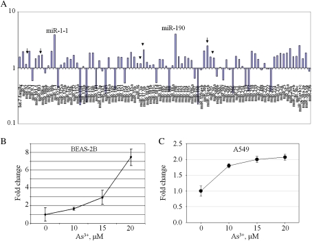
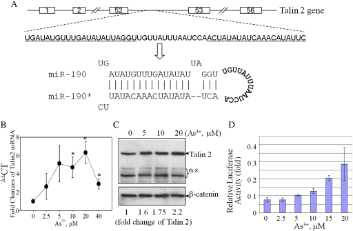
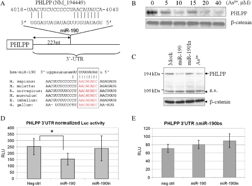
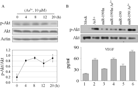
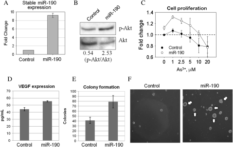
The role of trivalent arsenic (As(3+)) on the regulation of the recently identified noncoding small RNAs, mainly microRNAs, has not been explored so far. In the present study, we provide evidence showing that As(3+) is a potent inducer for the expression of miR-190 in human bronchial epithelial cells. The induction of miR-190 by As(3+) is concentration dependent and associated with the expression of the host gene of miR-190, talin 2, a gene encoding a high-molecular-weight cytoskeletal protein. The elevated level of miR-190 induced by As(3+) is capable of downregulating the translation of the PH domain leucine-rich repeat protein phosphatase (PHLPP), a negative regulator of Akt signaling. Such a downregulation is occurred through direct interaction of the miR-190 with the 3'-UTR region of the PHLPP mRNA, leading to a diminished PHLPP protein expression and consequently, an enhanced Akt activation and expression of vascular endothelial growth factor, an Akt-regulated protein. Overexpression of miR-190 itself is able to enhance proliferation and malignant transformation of the cells as determined by anchorage-independent growth of the cells in soft agar. Accordingly, the data presented suggest that induction of miR-190 is one of the key mechanisms in As(3+)-induced carcinogenesis.


Figures

FIG. 1.
FIG. 1.
As3+ regulates expression of miRNAs. (A) A real-time PCR-based miRNA array using Cancer miRNA Panel shows expression of miRNAs after As3+ exposure. Arrows indicate miRNAs in the miR-106-363 cluster; arrowheads indicate miRNAs in the miR-17-92 cluster. Data are representative of three experiments. (B) As3+ induces expression of miR-190. Real-time PCR was performed using total RNA extracted from the BEAS-2B cells treated with the indicated concentrations of As3+ for 12 h. Data are means ± SD of five experiments. (C) As3+ induces miR-190 in A549 cells. Cells were treated as in B. Data are means ± SD of three experiments.
FIG. 2.
FIG. 2.
Talin 2 expression in response to As3+. (A) Schematic diagram of the talin 2 gene and pre-miR-190 within the intron 52 region of the talin 2 gene. Underlined sequences indicate the sequences that form pre-miR-190 duplex through complementarily association of the inverted repeat region. The asterisk (*) indicates passage strand of the miR-190 duplex. (B) Real-time PCR analysis of talin 2 mRNA production in response to As3+. (C) Talin 2 protein was induced by a 12-h exposure of the cells with As3+ at the indicated concentrations. A loading control was made by Western blotting using antibody against β-catenin. n.s.: nonspecific bands. The bottom numbers show semiquantification of the talin 2 protein expression by densitometry. Data are representative of three experiments. (D) The talin 2 promoter reporter gene activity is activated by As3+ exposure. Data is normalized by calculating the ratio between Firefly luciferase activity versus Renilla luciferase activity.
FIG. 3.
FIG. 3.
miR-190 targets PHLPP. (A) Schematic diagram of the miR-190-binding site at the 3′-UTR of PHLPP mRNA. Lower panel shows alignment of the predicted miR-190-binding sites to 3-UTR of PHLPP from different species (Homo sapiens, Macaca mutatta, Rattus norvegicus, Mus musculus, Eguus caballus, and Gallus gallus). (B) Western blotting shows concentration-dependent reduction of the PHLPP protein in the cells treated with As3+. (C) Overexpression of miR-190 downregulates PHLPP. The BEAS-2B cells were mock transfected, or transfected with a miR-190, miR-190In, or treated with 20μM of As3+. The protein level of PHLPP was determined by Western blotting. (D) Overexpression of miR-190, but not miR-190In, downregulates the activity of the PHLPP 3′-UTR reporter gene. (E) Overexpression of miR-190 has no effect on the PHLPP 3′-UTR reporter without the miR-190-binding site.
FIG. 4.
FIG. 4.
Overexpression of miR-190 enhances As3+-induced Akt phosphorylation and VEGF generation. (A) BEAS-2B cells treated with 10μM As3+ for the indicated hours, and Akt phosphorylation at Ser473 was determined by Western blotting. The bottom panel shows relative levels of Akt phosphorylation as determined by the ratio of phospho-Akt versus nonphospho-Akt that were determined by densitometry scanning of three independent Western blotting experiments. (B) BEAS-2B cells were transfected with the miR-190 and miR-190In, respectively. Phosphorylation of Akt at Ser473 was analyzed by Western blot. The bottom panel shows VEGF expression following miRNA transfection and As3+ exposure by ELISA. Data shown is representative of three experiments.
FIG. 5.
FIG. 5.
Stable expression of miR-190 enhances cell proliferation and carcinogenic transformation. (A) The levels of miR-190 in the cells stably transfected with a control miRNA and miR-190 were determined by real-time PCR. (B) Levels of Akt activation in the stably transfected cells were determined by Western blotting using antibody against Ser473-phosphorylated Akt (top panel) or the nonphospho-Akt (bottom panel). The numbers at the bottom of the lower panel indicate ratios of phospho-Akt versus nonphospho-Akt. (C) The stably transfected BEAS-2B cells were cultured for 16 h followed by As3+ treatment with the indicated concentrations for an additional 12 h. Cell proliferation was determined as described in the “Experimental Procedures.” (D) VEGF levels were determined by ELISA in the stably transfected cells. (E) Quantification of the colony numbers of the stably transfected cells in soft agar. (F) Anchorage-independent growth of the stably transfected cells was determined by the colony formation in soft agar. White arrows indicate large colonies formed by the cells stably transfected with miR-190.

References

    1. Beezhold KJ, Castranova V, Chen F. Microprocessor of microRNAs: regulation and potential for therapeutic intervention. Mol. Cancer. 2010;9:134. - PMC - PubMed
    1. Calin GA, Liu CG, Sevignani C, Ferracin M, Felli N, Dumitru CD, Shimizu M, Cimmino A, Zupo S, Dono M, et al. MicroRNA profiling reveals distinct signatures in B cell chronic lymphocytic leukemias. Proc. Natl. Acad. Sci. U.S.A. 2004;101:11755–11760. - PMC - PubMed
    1. Camacho LM, Gutierrez M, Alarcon-Herrera MT, Villalba Mde L, Deng S. Occurrence and treatment of arsenic in groundwater and soil in northern Mexico and southwestern USA. Chemosphere. 2011;83:211–225. - PubMed
    1. Cao J, Tong C, Wu X, Lv J, Yang Z, Jin Y. Identification of conserved microRNAs in Bombyx mori (silkworm) and regulation of fibroin L chain production by microRNAs in heterologous system. Insect Biochem. Mol. Biol. 2008;38:1066–1071. - PubMed
    1. Cavigelli M, Li WW, Lin A, Su B, Yoshioka K, Karin M. The tumor promoter arsenite stimulates AP-1 activity by inhibiting a JNK phosphatase. EMBO J. 1996;15:6269–6279. - PMC - PubMed

Publication types

MeSH terms

LinkOut - more resources