. 2011;27(1):75-87.
doi: 10.3233/JAD-2011-101716.

Decreased proteolytic activity of the mitochondrial amyloid-β degrading enzyme, PreP peptidasome, in Alzheimer's disease brain mitochondria

Affiliations

Decreased proteolytic activity of the mitochondrial amyloid-β degrading enzyme, PreP peptidasome, in Alzheimer's disease brain mitochondria

Nyosha Alikhani et al. J Alzheimers Dis. 2011.

Abstract

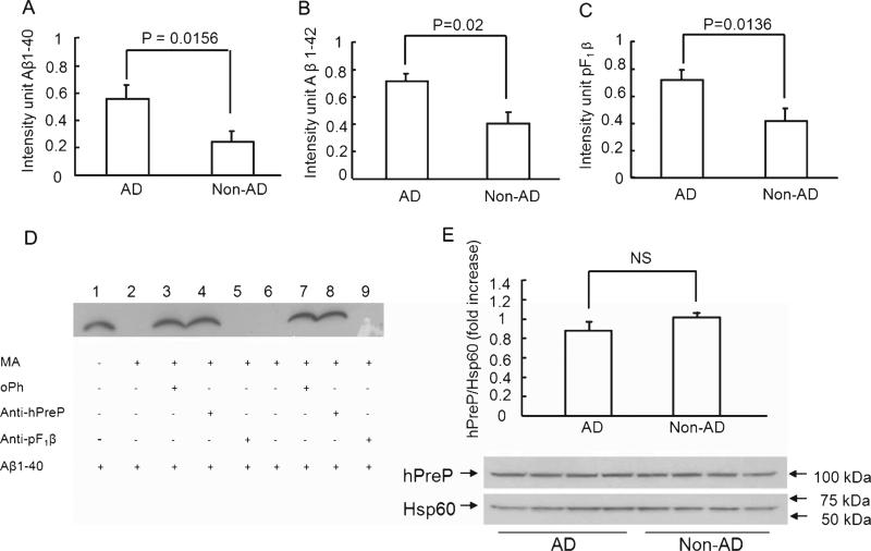
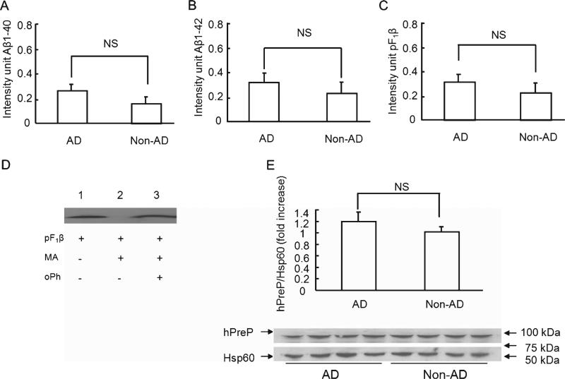
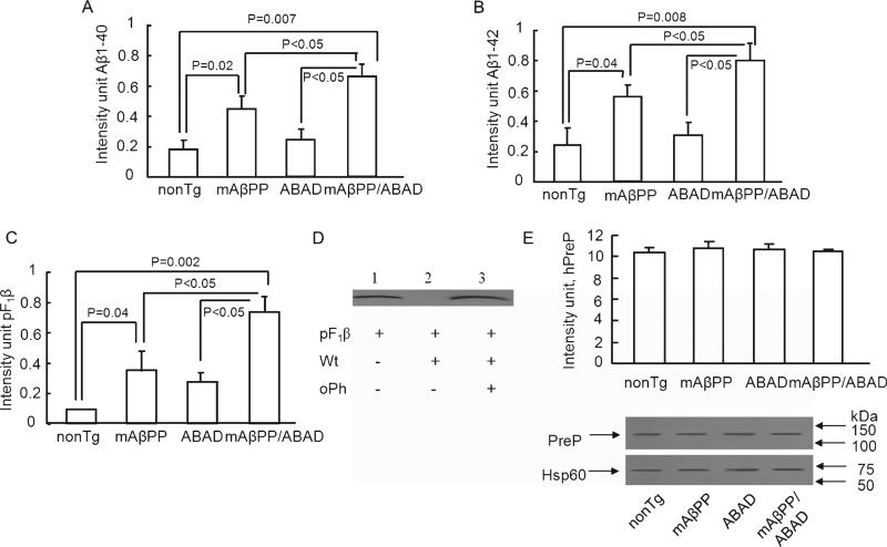
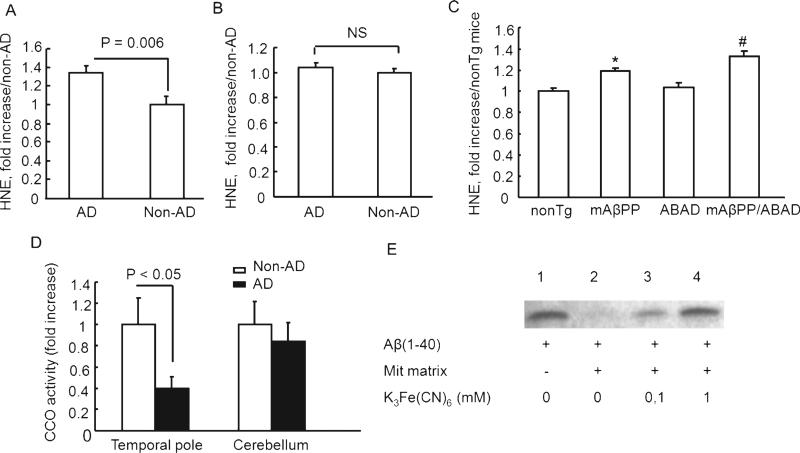
Accumulation of amyloid-β peptide (Aβ), the neurotoxic peptide implicated in the pathogenesis of Alzheimer's disease (AD), has been shown in brain mitochondria of AD patients and of AD transgenic mouse models. The presence of Aβ in mitochondria leads to free radical generation and neuronal stress. Recently, we identified the presequence protease, PreP, localized in the mitochondrial matrix in mammalian mitochondria as the novel mitochondrial Aβ-degrading enzyme. In the present study, we examined PreP activity in the mitochondrial matrix of the human brain's temporal lobe, an area of the brain highly susceptible to Aβ accumulation and reactive oxygen species (ROS) production. We found significantly lower hPreP activity in AD brains compared with non-AD age-matched controls. By contrast, in the cerebellum, a brain region typically spared from Aβ accumulation, there was no significant difference in hPreP activity when comparing AD samples to non-AD controls. We also found significantly reduced PreP activity in the mitochondrial matrix of AD transgenic mouse brains (Tg mAβPP and Tg mAβPP/ABAD) when compared to non-transgenic aged-matched mice. Furthermore, mitochondrial fractions isolated from AD brains and Tg mAβPP mice had higher levels of 4-hydroxynonenal, an oxidative product, as compared with those from non-AD and nonTg mice. Accordingly, activity of cytochrome c oxidase was significantly reduced in the AD mitochondria. These findings suggest that decreased PreP proteolytic activity, possibly due to enhanced ROS production, contributes to Aβ accumulation in mitochondria leading to the mitochondrial toxicity and neuronal death that is exacerbated in AD. Clearance of mitochondrial Aβ by PreP may thus be of importance in the pathology of AD.


Figures

Fig. 1
Fig. 1
Changes in hPreP activity/expression in brain mitochondria of AD temporal lobe. Degradation of biotin-Aβ (A, B) and F1β presequence peptide (C). hPreP proteins extracted from the cortical mitochondria of the indicated AD and non-AD brains (A, B) were incubated with biotin-labeled Aβ (0.25 μg), and then subjected to immunoblotting with Extravidin peroxidase conjugated IgG and detection with ECL to reveal immunoreactive biotin Aβ. C) For degradation of F1β substrate, mitochondrial hPreP proteins were incubated with pF1β followed by the immunoblot with antibody to pF1β. Densitometry of the combined immunoreactive Aβ (A, B) or F1β bands (C) using NIH Image program is shown. D) Determination of hPreP proteolytic activity and hPreP-dependent degradation of Aβ in brain mitochondrial fraction. Biotin-labeled Aβ was completely degraded by the isolated human mitochondrial matrix hPreP protein (MA-hPreP) (lanes 2 and 6). The oPh (ortho-phenantroline, an inhibitor of PreP proteolytic activity) blocked the degradation of biotin Aβ (lanes 3 and 7). The immuno-inactivation assay confirmed hPreP-induced degradation of Aβ. When isolated human mitochondrial matrix hPreP protein (MA-hPreP) was pre-incubated with antibody against hPreP, degradation of Aβ1-40 (lane 4 & 8) was almost completely inhibited, whereas incubation of pF1β presequence antibodies did not affect Aβ degradation (lanes 5 and 9). E) Immunoblotting of brain mitochondria from AD and non-AD brains for hPreP. The upper panel denotes representative immunoblots for human PreP and Hsp60. Hsp60 was used as a mitochondrial marker and protein loading controls. The lower panel shows densitometry of hPreP immunoreactive bands from the indicated brain mitochondria. NS: no statistical significance between these groups (p > 0.05). Mitochondria were isolated from 12 AD brains and 8 non-AD brains. All experiments were performed in triplicates.
Fig. 2
Fig. 2
hPreP activity/expression in the cerebellum of AD and non-AD brains. Mitochondrial hPreP extracted from cerebellum for degrading biotin Aβ40/42 (A,B) and pF1β (C). Densitometry of Aβ or pF1β immunoreactive bands are shown by diagram bars. D) Effect of hPreP activity on degrading of F1β presequence (pF1β). pF1β was completely degraded by mitochondrial matrix hPreP protein (MA-hPreP, no F1β immunreactive band in lane 2 versus F1β immunoreactive band in lane 1 without MA-hPreP). In the presence of oPh, MA-PreP was not able to degrade pF1β (lane 3 versus lane 2 without oPh). E) Immunoblotting of cortical mitochondria from AD and non-AD cerebellum for human PreP. NS: no significant difference. Mitochondria were isolated from 12 AD brains and 8 non-AD brains. All experiments were performed in triplicates.
Fig. 3
Fig. 3
Alterations of PreP activity/expression in transgenic AD mice. Cortical mitochondrial PreP extracted from 5-month-old indicated Tg mice degraded the biotin Aβ40 (A), Aβ42 (B), and pF1β (C), respectively. Densitometry of Aβ40/42 or pF1β immunoreactive bands was performed using NIH image program. The upper panels indicate the representative immunoblots for Aβ (A, B) and pF1β (D). In the presence of oPh, mitochondrial PreP was not able to degrade pF1β peptide (lane 3 vs. lane 2). E) Immunoblotting of cortical mitochondrial protein from the indicated Tg mice for PreP. Representative immunoblots for PreP and Hsp60 are shown in the upper panel. Hsp60 (mitochondrial marker) was used as protein loading control and mitochondrial rich fractions. n = 4–7 mice per group. All experiments were performed in triplicates.
Fig. 4
Fig. 4
PreP activity/expression in Tg AD mice at age of twelve months. The cortical mitochondrial PreP from twelve-months-old Tg mice degraded biotin Aβ40/42 (A,B) and pF1β (C) Densitometry of combined immunoreactive bands (Aβ or pF1β) from the indicated Tg mice. D) Immunoblotting of mitochondrial fractions from the indicated Tg mice for PreP or Hsp60. n = 4–5 mice per group. All experiments were performed in triplicates.
Fig. 5
Fig. 5
Age-dependent decreased PreP activity in Tg mAβPP mice. Cortical mitochondrial PreP was extracted from Tg mAβPP and nonTg mice at ages of five and twelve months, and then incubated with biotin Aβ40/42 (A, B) and pF1β (C), respectively. Densitomery of all immunoreactive bands for Aβ and pF1β is shown. n = 4–6 mice per group. All experiments were performed in triplicates.
Fig. 6
Fig. 6
A. HNE levels and cytochrome c oxidase activity in brain mitochondria. HNE levels in cortical mitochondria isolated from temporal lobe (A) and cerebellum (B) of AD and non-AD brains were measured by ELISA. n = 10–11 mice per group. C) HNE levels in cortical mitochondria from the indicated Tg mice were determined by ELISA. *p < 0.05 vs. nonTg mice, #p < 0.05 vs. nonTg and mAβPP mice. n = 8–9 mice per group. D) Cytochrome c oxidase activity in cortical mitochondria from temporal lobe and cerebellum of AD and non-AD subjects. E) Decreased hPreP activity in mitochondrial matrix in the presence of 0.1 mM and 1 mM K3Fe(CN)6 (lanes 3 and 4).

References

    1. Kimberly WT, LaVoie MJ, Ostaszewski BL, Ye W, Wolfe MS, Selkoe DJ. Gamma-secretase is a membrane protein complex comprised of presenilin, nicastrin, Aph-1, and Pen-2. Proc Natl Acad Sci U S A. 2003;100:6382–6387. - PMC - PubMed
    1. Selkoe DJ. Translating cell biology into therapeutic advances in Alzheimer's disease. Nature. 1999;399:A23–A31. - PubMed
    1. Masters CL, Simms G, Weinman NA, Multhaup G, McDonald BL, Beyreuther K. Amyloid plaque core protein in Alzheimer disease and Down syndrome. Proc Natl Acad Sci U S A. 1985;82:4245–4249. - PMC - PubMed
    1. Soderberg L, Bogdanovic N, Axelsson B, Winblad B, Naslund J, Tjernberg LO. Analysis of single Alzheimer solid plaque cores by laser capture microscopy and nano-electrospray/tandem mass spectrometry. Biochemistry. 2006;45:9849–9856. - PubMed
    1. Gouras GK, Almeida CG, Takahashi RH. Intraneuronal Abeta accumulation and origin of plaques in Alzheimer's disease. Neurobiol Aging. 2005;26:1235–1244. - PubMed

Publication types

MeSH terms