. 2011 Sep;19(9):1656-66.
doi: 10.1038/mt.2011.138. Epub 2011 Jul 12.

Protein transfer into human cells by VSV-G-induced nanovesicles

Affiliations

Protein transfer into human cells by VSV-G-induced nanovesicles

Philippe-Emmanuel Mangeot et al. Mol Ther. 2011 Sep.

Abstract

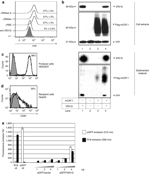
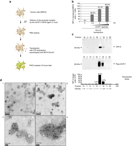
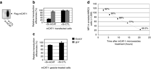
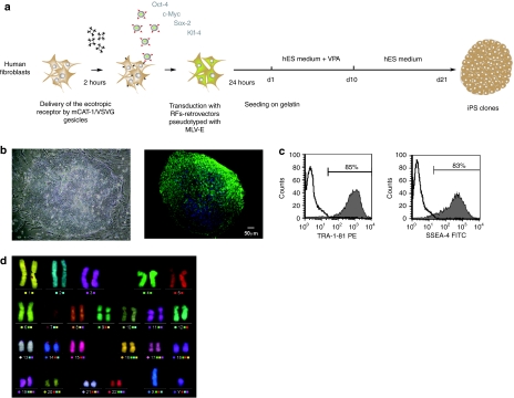
Identification of new techniques to express proteins into mammal cells is of particular interest for both research and medical purposes. The present study describes the use of engineered vesicles to deliver exogenous proteins into human cells. We show that overexpression of the spike glycoprotein of the vesicular stomatitis virus (VSV-G) in human cells induces the release of fusogenic vesicles named gesicles. Biochemical and functional studies revealed that gesicles incorporated proteins from producer cells and could deliver them to recipient cells. This protein-transduction method allows the direct transport of cytoplasmic, nuclear or surface proteins in target cells. This was demonstrated by showing that the TetR transactivator and the receptor for the murine leukemia virus (MLV) envelope [murine cationic amino acid transporter-1 (mCAT-1)] were efficiently delivered by gesicles in various cell types. We further shows that gesicle-mediated transfer of mCAT-1 confers to human fibroblasts a robust permissiveness to ecotropic vectors, allowing the generation of human-induced pluripotent stem cells in level 2 biosafety facilities. This highlights the great potential of mCAT-1 gesicles to increase the safety of experiments using retro/lentivectors. Besides this, gesicles is a versatile tool highly valuable for the nongenetic delivery of functions such as transcription factors or genome engineering agents.


Figures

Figure 1
Figure 1
Vesicular stomatitis virus (VSV-G) promotes the cell-release of a sedimentable agent capable of pseudotransduction. (a) FACS-analyzed pseudotransduction mediated by the VSVG/YFP agent. Two micrograms of sedimented material prepared from VSV-G/YFP cells were treated with RNAse A (10 µg/ml), DNAse I (50 U/ml) or with phosphate-buffered saline (PBS) before incubation with recipient HEK-293T cells. Material shedding from mock-transfected yellow fluorescence protein (YFP)-cells was similarly prepared and incubated with target cells, but failed at transferring YFP (w/o VSV-G). Histogram for nontreated cells is shown (NT). YFP histograms in gated populations and percentages of YFP+ cells are given for each condition. (b) Immunoblot analysis of the sedimented materials shedding from YFP-HEK-293T cells transfected with VSV-G (lane 1), mock-transfected (lane 2), flag-murine cationic amino acid transporter-1 (mCAT-1) (lane 3), flag-mCAT-1 and VSV-G (lane 4). Immunostaining of VSV-G, flag-mCAT-1, and YFP were performed. Each lane was loaded with 5 µg of protein as dosed by Bradford. Extracts from producer cells were similarly analyzed (upper panel). (c) CD81 expression on HEK-293T producer cells (open line). An isotypic control was used as a control staining (gray-filled histogram). (d) CD81 expression on HepG2 cells after incubation with a concentrated material derived from VSV-G-expressing producer cells (open line). Staining of nontreated HepG2 cell is shown (gray-filled histogram). (e) Fluorometric analysis of the VSV-G induced material produced from cells expressing enhanced green fluorescent protein (eGFP) and probed with a membrane tracer (R18). Producer cells were treated with octadecyl-Rhodamine B (R18) prior VSV-G transfection and sedimentation of the shedding material. Increasing amounts of the resulting material (eGFP/VSV-G) were analyzed by fluorometry for eGFP- and R18-emission. A preparation produced without VSV-G was also analyzed (eGFP/carrrier). eGFP-free gesicles loaded with R18 (R18 ctl) and R18-free gesicles-containing eGFP (eGFP ctl) were used as controls, showing the absence of overlapping between the two spectra. The experiments were performed in triplicate and the data represent means ± s.d.
Figure 2
Figure 2
Delivery of murine cationic amino acid transporter-1 (mCAT-1) in human cells by characterized gesicles. (a) Principle of the ecotropic transduction assay using simian immunodeficiency virus (SIV)-derived lentivectors pseudotyped with the murine leukemia virus (MLV)-E envelope (EcoLV). (b) Ecotropic transduction assay in human MRC-5 fibroblasts treated with increasing amounts of mCAT-1 gesicles. Seventy hours after transduction [multiplicity of infection (MOI) 40, 43 transducing units (TU)/pg P24], mean fluorescence intensity (MFIs) of gated populations and percentage of transduced cells were measured by FACS. The error bars represent the s.d. of triplicate MFI values. (c) Fractionation of the mCAT-1/vesicular stomatitis virus (VSV-G) pellet on a iodixanol gradient. After ultracentrifugation, fractions were collected and analyzed by immunoblotting. VSV-G and mCAT-1 proteins were immunodetected mainly in fractions 17 and 18. mCAT-1 transfer activity of each fraction was measured by an ecotropic transduction using HEK-293T target cells. Percentages of transduced cells and MFI are given. Nonrepresented fractions 1–11 revealed no staining neither receptor-transfer activity. Density of each fraction is indicated. (d) Electron microscopy observation of mCAT-1 gesicles after immunostaining with a control antibody (panel 1) or an anti-flag antibody recognizing flag-mCAT-1 (panels 2–4). A higher magnification of the upper-left region of panel 2 (open square) is shown in panel 3.
Figure 3
Figure 3
Gesicles delivered murine cationic amino acid transporter-1 (mCAT-1) as a functional protein. (a) Validation of the mCAT-1 small interfering RNA (siRNA). Flag mCAT-1 immunodetection was performed in cell extracts derived from HEK-293T transfected with mCAT-1 in addition with the mCAT-1 siRNA (Si-mCAT lane) or a control siRNA (Si-CTL lane). Ten microgram of each protein extract were loaded per lane. (b) Ecotropic transduction assay in HEK-293T cells after delivery of the receptor by mCAT-1 transfection. EcoLV (black bars) and gLV (gray bars) stand for ecotropic lentivector transduction and pantropic lentivector transduction [vesicular stomatitis virus (VSV-G) pseudotyped], respectively. EcoLV transduction was impaired by the mCAT-1 siRNA. (c) Ecotropic transduction assay in HEK-293T cells after delivery of the receptor by mCAT-1 gesicles. The mCAT-1 siRNA poorly affected the receptor activity when mCAT-1 was delivered by gesicles. (d) Residence time of mCAT-1 in cells treated by gesicles. Mitomycin arrested MRC-5 cells (105 cells in a 12-well plate) were treated with 2 µg of mCAT-1 gesicles and transduced with an ecotropic yellow fluorescence protein (YFP) lentivector [multiplicity of infection (MOI) 20] 3 hours, 5 hours, 9 hours, 14 hours, and 20 hours post-treatment. Transduction efficiencies were measured 72 hours later by FACS. Mean fluorescence intensity (MFIs) of total living populations and percentages of transduced MRC-5 cells are given for each condition. Error bars indicates s.d. (n = 3).
Figure 4
Figure 4
iPS generation assisted with murine cationic amino acid transporter-1 (mCAT-1) gesicles. (a) Reprogramming protocol. (b) Left: Phase contrast representation of an iPSC clone obtained from ecotropic retroviral reprogramming. Right: Immunostaining of the established iPSC clone. Green SSEA-4 staining. Nuclei appear in blue (DAPI staining). (c) FACS analysis of iPSC clone after immunostaining with pluripotency markers. (d) mFISH analysis showing normal karyotype at passage 41.
Figure 5
Figure 5
Gesicle-mediated delivery of TetR-regulators in human-cells. (a) Schematic representation of constructs encoding for TetR-regulators. Expression is driven by the human cytomegalovirus early promoter (hCMV) upstream the rabbit b-globin intron (i), and the rabbit b-globin polyadenylation signal (pA). Engineered tTA sequences incorporating a linker (L) and the C-terminal farnesyl motif of H-RAS are depicted (f). (b) Functional validation of the pptTA and pptTA-Lf constructs. (Up): Low amounts of both plasmids (0.1 µg/105 cells) were transfected in a reporter cell line-expressing GFP under the control of the tetO promoter (tetOG cells). Percentages of transfected cells and fluorescence intensities were measured 24 hours later. (Bottom): Immunodetection of the two proteins in cell-extracts. Ten microgram of total protein were loaded per lane. Mock stands for control mock-transfection. (c) Transfer of TetR-activators by gesicles. (Up): Activation of green fluorescent protein (GFP)-transcription in tetOG cells 48 hours after treatment with vesicles derived from producer cells transfected with pptTA or pptTA-Lf with or without vesicular stomatitis virus (VSV-G). (10 µg of gesicles/105 cells). Mock stands for non treated reporter cells. Percentages of GFP+ cells targeted by gesicles are given. (Bottom): Western blot analysis of resulting sedimented vesicles (d) Dose-dependent activation of GFP-transcription in tetOG cells after treatment with tTA-Lf gesicles. Mean fluorescence intensity (MFIs) of recipient cells and percentages of GFP+ cells were measured by FACS 48 hours after treatment. Doses of gesicles are given in µg. (e) Downregulation of transgene expression in human cells by tTRKrab gesicles. A leaky tetO-GFP cell line was generated which emits a fluorescence signal without induction (left bar). 95% of this reporter cell line was GFP+ as compared with parental HEK-293T. GFP downregulation was analyzed by FACS 60 hours after gesicle treatment. The data represent means ± s.d. (n = 3).

Comment in

References

    1. Wagstaff KM., and, Jans DA. Protein transduction: cell penetrating peptides and their therapeutic applications. Curr Med Chem. 2006;13:1371–1387. - PubMed
    1. Mäe M., and, Langel U. Cell-penetrating peptides as vectors for peptide, protein and oligonucleotide delivery. Curr Opin Pharmacol. 2006;6:509–514. - PubMed
    1. Liguori L., and, Lenormand JL. Production of recombinant proteoliposomes for therapeutic uses. Meth Enzymol. 2009;465:209–223. - PubMed
    1. Xu YF, Zhang YQ, Xu XM., and, Song GX. Papillomavirus virus-like particles as vehicles for the delivery of epitopes or genes. Arch Virol. 2006;151:2133–2148. - PubMed
    1. Temchura VV, Tenbusch M, Nchinda G, Nabi G, Tippler B, Zelenyuk M.et al. (2008Enhancement of immunostimulatory properties of exosomal vaccines by incorporation of fusion-competent G protein of vesicular stomatitis virus Vaccine 263662–3672. - PMC - PubMed

Publication types

MeSH terms